• 文献检索
  • 文档翻译
  • 深度研究
  • 学术资讯
  • Suppr Zotero 插件Zotero 插件
  • 邀请有礼
  • 套餐&价格
  • 历史记录
应用&插件
Suppr Zotero 插件Zotero 插件浏览器插件Mac 客户端Windows 客户端微信小程序
定价
高级版会员购买积分包购买API积分包
服务
文献检索文档翻译深度研究API 文档MCP 服务
关于我们
关于 Suppr公司介绍联系我们用户协议隐私条款
关注我们

Suppr 超能文献

核心技术专利:CN118964589B侵权必究
粤ICP备2023148730 号-1Suppr @ 2026

文献检索

告别复杂PubMed语法,用中文像聊天一样搜索,搜遍4000万医学文献。AI智能推荐,让科研检索更轻松。

立即免费搜索

文件翻译

保留排版,准确专业,支持PDF/Word/PPT等文件格式,支持 12+语言互译。

免费翻译文档

深度研究

AI帮你快速写综述,25分钟生成高质量综述,智能提取关键信息,辅助科研写作。

立即免费体验

骨桥蛋白表达缺失可减少 HSV-1 诱导的角膜混浊。

Loss of Osteopontin Expression Reduces HSV-1-Induced Corneal Opacity.

机构信息

Dean McGee Eye Institute, Department of Ophthalmology, University of Oklahoma, Oklahoma City, Oklahoma, United States.

Department of Microbiology and Immunology, University of Oklahoma Health Sciences Center, Oklahoma City, Oklahoma, United States.

出版信息

Invest Ophthalmol Vis Sci. 2020 Aug 3;61(10):24. doi: 10.1167/iovs.61.10.24.

DOI:10.1167/iovs.61.10.24
PMID:32785676
原文链接:https://pmc.ncbi.nlm.nih.gov/articles/PMC7441335/
Abstract

PURPOSE

Corneal opacity and neovascularization (NV) are often described as outcomes of severe herpes simplex virus type 1 (HSV-1) infection. The current study investigated the role of colony-stimulating factor 1 receptor (CSF1R)+ cells and soluble factors in the progression of HSV-1-induced corneal NV and opacity.

METHODS

MaFIA mice were infected with 500 plaque-forming units of HSV-1 in the cornea following scarification. From day 10 to day 13 post-infection (pi), mice were treated with 40 µg/day of AP20187 (macrophage ablation) or vehicle intraperitoneally. For osteopontin (OPN) neutralization experiments, C57BL/6 mice were infected as above and treated with 2 µg of goat anti-mouse OPN or isotypic control IgG subconjunctivally every 2 days from day 4 to day 12 pi. Mice were euthanized on day 14 pi, and tissue was processed for immunohistochemistry to quantify NV and opacity by confocal microscopy and absorbance or detection of pro- and anti-angiogenic and inflammatory factors and cells by suspension array analysis and flow cytometry, respectively.

RESULTS

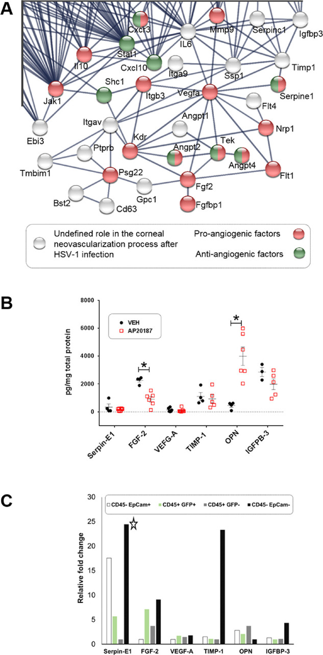
In the absence of CSF1R+ cells, HSV-1-induced blood and lymphatic vessel growth was muted. These results correlated with a loss in fibroblast growth factor type 2 (FGF-2) and an increase in OPN expression in the infected cornea. However, a reduction in OPN expression in mice did not alter corneal NV but significantly reduced opacity.

CONCLUSIONS

Our data suggest that CSF1R+ cell depletion results in a significant reduction in HSV-1-induced corneal NV that correlates with the loss of FGF-2 expression. A reduction in OPN expression was aligned with a significant drop in opacity associated with reduced corneal collagen disruption.

摘要

目的

角膜混浊和新生血管(NV)常被描述为单纯疱疹病毒 1 型(HSV-1)严重感染的结果。本研究探讨了集落刺激因子 1 受体(CSF1R)+细胞和可溶性因子在 HSV-1 诱导的角膜 NV 和混浊进展中的作用。

方法

MaFIA 小鼠经划痕后在角膜中感染 500 噬菌斑形成单位的 HSV-1。感染后第 10 天至第 13 天,小鼠每天腹膜内给予 40μg/天的 AP20187(巨噬细胞消融)或载体。为了进行骨桥蛋白(OPN)中和实验,如上所述感染 C57BL/6 小鼠,并从感染后第 4 天至第 12 天每 2 天通过结膜下给予 2μg 山羊抗小鼠 OPN 或同种型对照 IgG。感染后第 14 天处死小鼠,通过共聚焦显微镜对 NV 和混浊进行免疫组织化学量化,并通过悬浮阵列分析和流式细胞术分别检测前血管生成、抗血管生成和炎症因子和细胞。

结果

在缺乏 CSF1R+细胞的情况下,HSV-1 诱导的血液和淋巴管生长减弱。这些结果与感染角膜中成纤维细胞生长因子 2(FGF-2)的丧失和 OPN 表达的增加相关。然而,在小鼠中降低 OPN 表达并没有改变角膜 NV,但显著降低了混浊度。

结论

我们的数据表明,CSF1R+细胞耗竭导致 HSV-1 诱导的角膜 NV 显著减少,这与 FGF-2 表达的丧失相关。OPN 表达的减少与与角膜胶原破坏减少相关的显著混浊度下降相关。

https://cdn.ncbi.nlm.nih.gov/pmc/blobs/ec1d/7441335/f9b58c487308/iovs-61-10-24-f007.jpg
https://cdn.ncbi.nlm.nih.gov/pmc/blobs/ec1d/7441335/31cc4902628f/iovs-61-10-24-f001.jpg
https://cdn.ncbi.nlm.nih.gov/pmc/blobs/ec1d/7441335/74a6d785d0ac/iovs-61-10-24-f002.jpg
https://cdn.ncbi.nlm.nih.gov/pmc/blobs/ec1d/7441335/021f72c31070/iovs-61-10-24-f003.jpg
https://cdn.ncbi.nlm.nih.gov/pmc/blobs/ec1d/7441335/dba2170310c7/iovs-61-10-24-f004.jpg
https://cdn.ncbi.nlm.nih.gov/pmc/blobs/ec1d/7441335/9b0475460a43/iovs-61-10-24-f005.jpg
https://cdn.ncbi.nlm.nih.gov/pmc/blobs/ec1d/7441335/acfb36c30bc8/iovs-61-10-24-f006.jpg
https://cdn.ncbi.nlm.nih.gov/pmc/blobs/ec1d/7441335/f9b58c487308/iovs-61-10-24-f007.jpg

相似文献

1
Loss of Osteopontin Expression Reduces HSV-1-Induced Corneal Opacity.骨桥蛋白表达缺失可减少 HSV-1 诱导的角膜混浊。
Invest Ophthalmol Vis Sci. 2020 Aug 3;61(10):24. doi: 10.1167/iovs.61.10.24.
2
Osteopontin contributes to virus resistance associated with type I IFN expression, activation of downstream ifn-inducible effector genes, and CCR2CD115CD206 macrophage infiltration following ocular HSV-1 infection of mice.骨桥蛋白有助于 I 型干扰素表达相关的病毒抵抗、下游 IFN 诱导效应基因的激活以及 CCR2+CD115+CD206+巨噬细胞浸润,这是在小鼠眼表单纯疱疹病毒-1 感染后观察到的现象。
Front Immunol. 2023 Jan 4;13:1028341. doi: 10.3389/fimmu.2022.1028341. eCollection 2022.
3
Effect of human apolipoprotein E genotype on the pathogenesis of experimental ocular HSV-1.人类载脂蛋白E基因型对实验性眼部单纯疱疹病毒1型发病机制的影响。
Exp Eye Res. 2008 Aug;87(2):122-30. doi: 10.1016/j.exer.2008.05.007. Epub 2008 May 18.
4
Colony Stimulating Factor-1 Receptor Expressing Cells Infiltrating the Cornea Control Corneal Nerve Degeneration in Response to HSV-1 Infection.表达集落刺激因子-1受体的细胞浸润角膜,可控制单纯疱疹病毒1型感染后的角膜神经变性。
Invest Ophthalmol Vis Sci. 2017 Sep 1;58(11):4670-4682. doi: 10.1167/iovs.17-22159.
5
Linkage of IL-6 with neutrophil chemoattractant expression in virus-induced ocular inflammation.白细胞介素-6在病毒诱导的眼部炎症中与中性粒细胞趋化因子表达的关联。
Invest Ophthalmol Vis Sci. 2002 Mar;43(3):737-43.
6
Exposure Stress Induces Reversible Corneal Graft Opacity in Recipients With Herpes Simplex Virus-1 Infections.暴露应激在单纯疱疹病毒-1感染的受体中诱导可逆性角膜移植混浊。
Invest Ophthalmol Vis Sci. 2017 Jan 1;58(1):35-41. doi: 10.1167/iovs.16-19673.
7
The use of human cornea organotypic cultures to study herpes simplex virus type 1 (HSV-1)-induced inflammation.利用人角膜器官型培养物研究单纯疱疹病毒1型(HSV-1)诱导的炎症。
Graefes Arch Clin Exp Ophthalmol. 2015 Oct;253(10):1721-8. doi: 10.1007/s00417-015-3073-4. Epub 2015 Jun 6.
8
Fibroblast growth factor-2 drives and maintains progressive corneal neovascularization following HSV-1 infection.成纤维细胞生长因子-2 驱动并维持 HSV-1 感染后的进行性角膜新生血管化。
Mucosal Immunol. 2018 Jan;11(1):172-185. doi: 10.1038/mi.2017.26. Epub 2017 Apr 5.
9
Herpes simplex virus-1 KOS-63 strain is virulent and causes titer-dependent corneal nerve damage and keratitis.单纯疱疹病毒-1 KOS-63 株是一种毒力很强的病毒,会导致剂量依赖性角膜神经损伤和角膜炎。
Sci Rep. 2021 Feb 19;11(1):4267. doi: 10.1038/s41598-021-83412-9.
10
CCR6-Positive γδ T Cells Provide Protection Against Intracorneal HSV-1 Infection.CCR6+γδ T 细胞为角膜内 HSV-1 感染提供保护。
Invest Ophthalmol Vis Sci. 2019 Sep 3;60(12):3952-3962. doi: 10.1167/iovs.19-27810.

引用本文的文献

1
Early expression of osteopontin glycoprotein on the ocular surface and in tear fluid contributes to ocular surface diseases in type 2 diabetic mice.骨桥蛋白糖蛋白在眼部表面和泪液中的早期表达有助于 2 型糖尿病小鼠的眼表面疾病。
PLoS One. 2024 Oct 31;19(10):e0313027. doi: 10.1371/journal.pone.0313027. eCollection 2024.
2
Herpes simplex keratitis: A brief clinical overview.单纯疱疹性角膜炎:简要临床概述。
World J Virol. 2024 Mar 25;13(1):89934. doi: 10.5501/wjv.v13.i1.89934.
3
The miR-183/96/182 cluster is a checkpoint for resident immune cells and shapes the cellular landscape of the cornea.

本文引用的文献

1
Progress in Research on the Role of FGF in the Formation and Treatment of Corneal Neovascularization.成纤维细胞生长因子在角膜新生血管形成与治疗中作用的研究进展
Front Pharmacol. 2020 Feb 25;11:111. doi: 10.3389/fphar.2020.00111. eCollection 2020.
2
Role of Insulin-Like Growth Factor Binding Protein-3 in the Pathogenesis of Herpes Stromal Keratitis.胰岛素样生长因子结合蛋白-3 在疱疹性基质性角膜炎发病机制中的作用。
Invest Ophthalmol Vis Sci. 2020 Feb 7;61(2):46. doi: 10.1167/iovs.61.2.46.
3
The role of extracellular matrix in mouse and human corneal neovascularization.
miR-183/96/182 簇是常驻免疫细胞的检查点,可塑造角膜的细胞景观。
Ocul Surf. 2023 Oct;30:17-41. doi: 10.1016/j.jtos.2023.07.012. Epub 2023 Aug 2.
4
The Chemistry and Biology of Collagen Hybridization.胶原杂交的化学与生物学。
J Am Chem Soc. 2023 May 24;145(20):10901-10916. doi: 10.1021/jacs.3c00713. Epub 2023 May 9.
5
Osteopontin contributes to virus resistance associated with type I IFN expression, activation of downstream ifn-inducible effector genes, and CCR2CD115CD206 macrophage infiltration following ocular HSV-1 infection of mice.骨桥蛋白有助于 I 型干扰素表达相关的病毒抵抗、下游 IFN 诱导效应基因的激活以及 CCR2+CD115+CD206+巨噬细胞浸润,这是在小鼠眼表单纯疱疹病毒-1 感染后观察到的现象。
Front Immunol. 2023 Jan 4;13:1028341. doi: 10.3389/fimmu.2022.1028341. eCollection 2022.
6
HSV-1 0∆NLS vaccine elicits a robust B lymphocyte response and preserves vision without HSV-1 glycoprotein M or thymidine kinase recognition.HSV-1 ∆NLS 疫苗可引起强烈的 B 淋巴细胞反应,并在不识别 HSV-1 糖蛋白 M 或胸苷激酶的情况下保留视力。
Sci Rep. 2022 Sep 23;12(1):15920. doi: 10.1038/s41598-022-20180-0.
7
Lymphatic Vessel Regression and Its Therapeutic Applications: Learning From Principles of Blood Vessel Regression.淋巴管消退及其治疗应用:从血管消退原理中学习
Front Physiol. 2022 Mar 22;13:846936. doi: 10.3389/fphys.2022.846936. eCollection 2022.
8
Tripartite-Motif 21 (TRIM21) Deficiency Results in a Modest Loss of Herpes Simplex Virus (HSV)-1 Surveillance in the Trigeminal Ganglia Following Cornea Infection.三基序蛋白 21(TRIM21)缺陷导致角膜感染后三叉神经节中单纯疱疹病毒 1(HSV-1)监测适度丢失。
Viruses. 2022 Mar 12;14(3):589. doi: 10.3390/v14030589.
9
The Durability of Vaccine Efficacy against Ocular HSV-1 Infection Using ICP0 Mutants 0∆NLS and 0∆RING Is Lost over Time.使用ICP0突变体0∆NLS和0∆RING的疫苗对眼部单纯疱疹病毒1型感染的效力持久性会随时间丧失。
Pathogens. 2021 Nov 12;10(11):1470. doi: 10.3390/pathogens10111470.
10
Lack of neonatal Fc receptor does not diminish the efficacy of the HSV-1 0ΔNLS vaccine against ocular HSV-1 challenge.缺乏新生 Fc 受体并不会降低 HSV-1 0ΔNLS 疫苗对眼部 HSV-1 挑战的疗效。
Vaccine. 2021 Apr 28;39(18):2526-2536. doi: 10.1016/j.vaccine.2021.03.075. Epub 2021 Apr 1.
细胞外基质在小鼠和人眼角膜新生血管中的作用。
Sci Rep. 2019 Oct 3;9(1):14272. doi: 10.1038/s41598-019-50718-8.
4
Methods for Assessing Corneal Opacity.评估角膜混浊的方法。
Semin Ophthalmol. 2019;34(4):205-210. doi: 10.1080/08820538.2019.1620796. Epub 2019 Jun 12.
5
Modulation of Corneal FAK/PI3K/Akt Signaling Expression and of Metalloproteinase-2 and Metalloproteinase-9 during the Development of Herpes Simplex Keratitis.调节单纯疱疹性角膜炎发展过程中角膜 FAK/PI3K/Akt 信号表达及金属蛋白酶-2 和金属蛋白酶-9
Biomed Res Int. 2019 Apr 2;2019:4143981. doi: 10.1155/2019/4143981. eCollection 2019.
6
STRING v11: protein-protein association networks with increased coverage, supporting functional discovery in genome-wide experimental datasets.STRING v11:具有增强覆盖范围的蛋白质-蛋白质相互作用网络,支持在全基因组实验数据集的功能发现。
Nucleic Acids Res. 2019 Jan 8;47(D1):D607-D613. doi: 10.1093/nar/gky1131.
7
The mouse autonomic nervous system modulates inflammation and epithelial renewal after corneal abrasion through the activation of distinct local macrophages.小鼠自主神经系统通过激活不同的局部巨噬细胞调节角膜擦伤后的炎症和上皮更新。
Mucosal Immunol. 2018 Sep;11(5):1496-1511. doi: 10.1038/s41385-018-0031-6. Epub 2018 Jul 9.
8
CD206-positive myeloid cells bind galectin-9 and promote a tumor-supportive microenvironment.CD206 阳性髓系细胞结合半乳糖凝集素-9 并促进肿瘤支持性微环境。
J Pathol. 2018 Aug;245(4):468-477. doi: 10.1002/path.5093. Epub 2018 Jun 28.
9
PD-L1/B7-H1 Inhibits Viral Clearance by Macrophages in HSV-1-Infected Corneas.PD-L1/B7-H1 抑制 HSV-1 感染角膜中的巨噬细胞清除病毒。
J Immunol. 2018 Jun 1;200(11):3711-3719. doi: 10.4049/jimmunol.1700417. Epub 2018 Apr 18.
10
Corneal Epithelial Cells Exhibit Myeloid Characteristics and Present Antigen via MHC Class II.角膜上皮细胞表现出髓样细胞特征,并通过 MHC Ⅱ类分子呈递抗原。
Invest Ophthalmol Vis Sci. 2018 Mar 1;59(3):1512-1522. doi: 10.1167/iovs.17-23279.