Chucair-Elliott Ana J, Gurung Hem R, Carr Meghan M, Carr Daniel J J
Department of Ophthalmology, University of Oklahoma Health Sciences Center, Oklahoma City, Oklahoma, United States.
Microbiology and Immunology, University of Oklahoma Health Sciences Center, Oklahoma City, Oklahoma, United States.
Invest Ophthalmol Vis Sci. 2017 Sep 1;58(11):4670-4682. doi: 10.1167/iovs.17-22159.
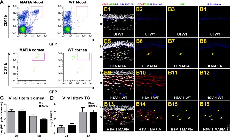
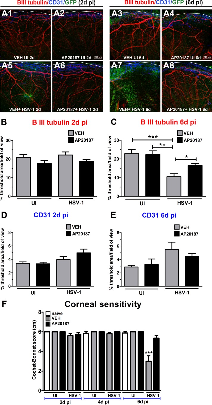
Herpes simplex virus type-1 (HSV-1) is a leading cause of neurotrophic keratitis, characterized by decreased or absent corneal sensation due to damage to the sensory corneal innervation. We previously reported the elicited immune response to infection contributes to the mechanism of corneal nerve regression/damage during acute HSV-1 infection. Our aim is to further establish the involvement of infiltrated macrophages in the mechanism of nerve loss upon infection.
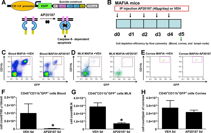
Macrophage Fas-Induced Apoptosis (MAFIA) transgenic C57BL/6 mice were systemically treated with AP20187 dimerizer or vehicle (VEH), and their corneas, lymph nodes, and blood were assessed for CD45+CD11b+GFP+ cell depletion by flow cytometry (FC). Mice were ocularly infected with HSV-1 or left uninfected. At 2, 4, and/or 6 days post infection (PI), corneas were assessed for sensitivity and harvested for FC, nerve structure by immunohistochemistry, viral content by plaque assay, soluble factor content by suspension array, and activation of signaling pathways by Western blot analysis. C57BL6 mice were used to compare to the MAFIA mouse model.
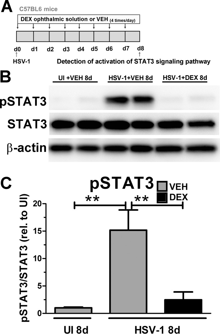
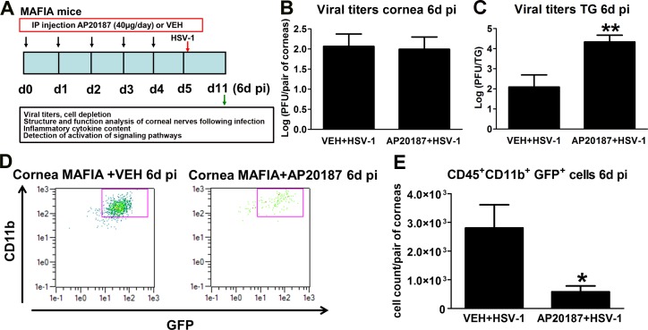
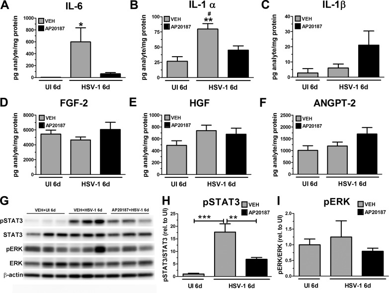
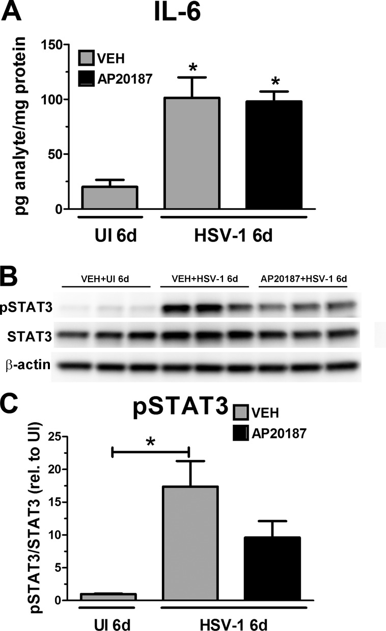
MAFIA mice treated with AP20187 had efficient depletion of CD45+CD11b+GFP+ cells in the tissues analyzed. The reduction of CD45+CD11b+GFP+ cells recruited to the infected corneas of AP20187-treated mice correlated with preservation of corneal nerve structure and function, decreased protein concentration of inflammatory cytokines, and decreased STAT3 activation despite no changes in viral content in the cornea compared to VEH-treated animals.
Our results suggest infiltrated macrophages are early effectors in the nerve regression following HSV-1 infection. We propose the neurodegeneration mechanism involves macrophages, local up-regulation of IL-6, and activation of STAT3.
单纯疱疹病毒1型(HSV-1)是神经营养性角膜炎的主要病因,其特征是由于角膜感觉神经支配受损导致角膜感觉减退或缺失。我们之前报道,感染引发的免疫反应参与了急性HSV-1感染期间角膜神经退化/损伤的机制。我们的目的是进一步确定浸润性巨噬细胞在感染后神经损伤机制中的作用。
用AP20187二聚体或载体(VEH)对巨噬细胞Fas诱导凋亡(MAFIA)转基因C57BL/6小鼠进行全身治疗,通过流式细胞术(FC)评估其角膜、淋巴结和血液中CD45+CD11b+GFP+细胞的清除情况。小鼠眼部感染HSV-1或不感染。在感染后(PI)2、4和/或6天,评估角膜的敏感性,并采集角膜用于FC分析、通过免疫组织化学评估神经结构、通过空斑试验评估病毒含量、通过悬浮阵列评估可溶性因子含量以及通过蛋白质印迹分析评估信号通路的激活情况。使用C57BL6小鼠与MAFIA小鼠模型进行比较。
用AP20187治疗的MAFIA小鼠在所分析的组织中有效清除了CD45+CD11b+GFP+细胞。与载体处理的动物相比,招募到AP20187处理小鼠感染角膜中的CD45+CD11b+GFP+细胞减少,这与角膜神经结构和功能的保留、炎性细胞因子蛋白浓度降低以及STAT3激活减少相关,尽管角膜中的病毒含量没有变化。
我们的结果表明,浸润性巨噬细胞是HSV-1感染后神经退化的早期效应细胞。我们提出神经退行性变机制涉及巨噬细胞、IL-6的局部上调和STAT3的激活。