Department of Cardiology, Leipzig University Hospital, Liebigstr. 20, 04103, Leipzig, Germany.
Department of Internal Medicine I, Martha-Maria Hospital Halle-Dölau, Röntgenstr. 1, 06129, Halle (Saale), Germany.
Clin Res Cardiol. 2020 Dec;109(12):1549-1566. doi: 10.1007/s00392-020-01727-5. Epub 2020 Aug 14.
Myocardial involvement induced by SARS-CoV-2 infection might be important for long-term prognosis. The aim of this observational study was to characterize the myocardial effects during SARS-CoV-2 infections by echocardiography.
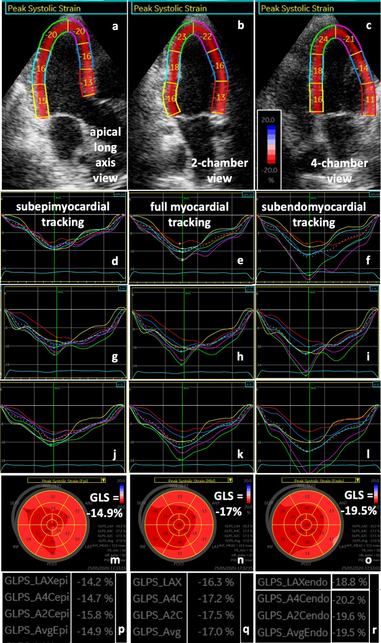
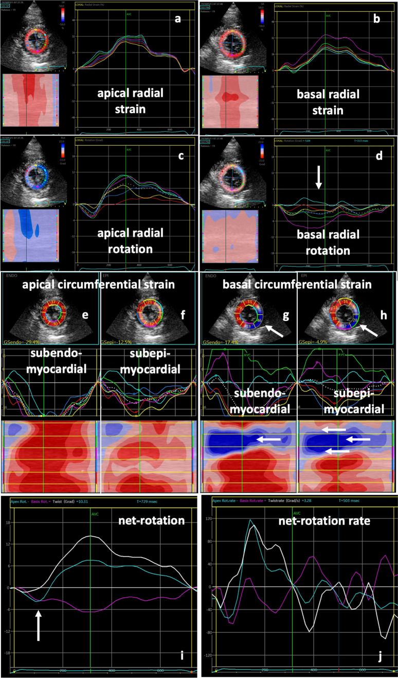
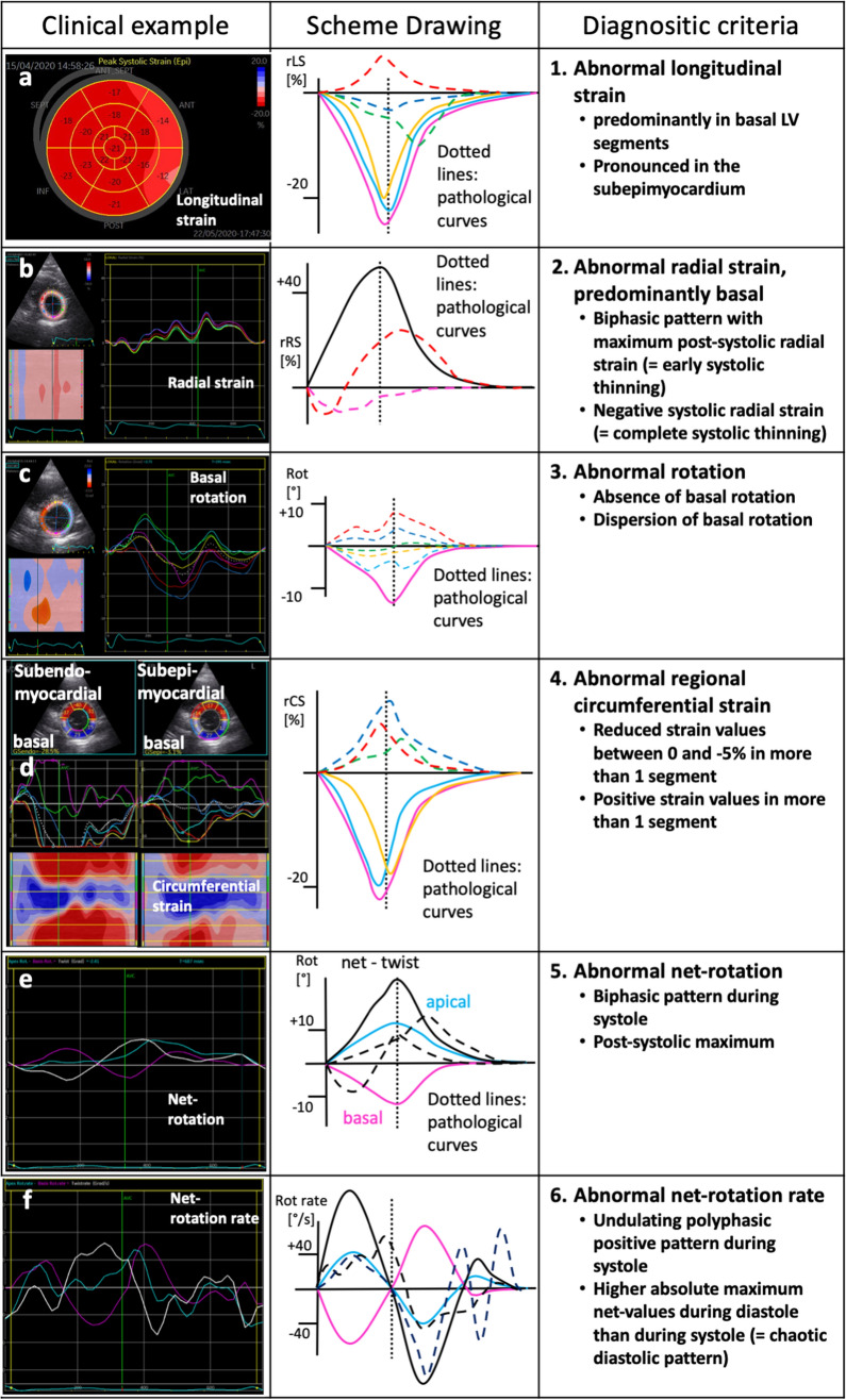
An extended echocardiographic image acquisition protocol was performed in 18 patients with SARS-CoV-2 infection assessing LV longitudinal, radial, and circumferential deformation including rotation, twist, and untwisting. Furthermore, LV deformation was analyzed in an age-matched control group of healthy individuals (n = 20). The most prevalent finding was a reduced longitudinal strain observed predominantly in more than one basal LV segment (n = 10/14 patients, 71%). This pattern reminded of a "reverse tako-tsubo" morphology that is not typical for other viral myocarditis. Additional findings included a biphasic pattern with maximum post-systolic or negative regional radial strain predominantly basal (n = 5/14 patients, 36%); the absence or dispersion of basal LV rotation (n = 6/14 patients, 43%); a reduced or positive regional circumferential strain in more than one segment (n = 7/14 patients, 50%); a net rotation showing late post-systolic twist or biphasic pattern (n = 8/14 patients, 57%); a net rotation showing polyphasic pattern and/or higher maximum net values during diastole (n = 8/14 patients, 57%).
Myocardial involvement due to SARS-CoV-2-infection was highly prevalent in the present cohort-even in patients with mild symptoms. It appears to be characterized by specific speckle tracking deformation abnormalities in the basal LV segments. These data set the stage to prospectively test whether these parameters are helpful for risk stratification and for the long-term follow-up of these patients.
由 SARS-CoV-2 感染引起的心肌受累可能对长期预后很重要。本观察性研究的目的是通过超声心动图来描述 SARS-CoV-2 感染期间的心肌效应。
对 18 例 SARS-CoV-2 感染患者进行了扩展的超声心动图图像采集方案,评估了左心室纵向、径向和周向变形,包括旋转、扭转和解旋。此外,还对 20 名健康个体的年龄匹配对照组进行了左心室变形分析。最常见的发现是观察到的纵向应变减少,主要发生在超过一个基底节段(n=10/14 例,71%)。这种模式让人联想到一种“反向心尖球囊样综合征”形态,这与其他病毒性心肌炎不同。其他发现包括收缩后或最大负性区域径向应变的双相模式,主要发生在基底节段(n=5/14 例,36%);基底节段左心室旋转的缺失或分散(n=6/14 例,43%);超过一个节段的区域性周向应变减少或阳性(n=7/14 例,50%);表现为收缩后晚期扭转或双相模式的净旋转(n=8/14 例,57%);表现为多相模式和/或舒张期最大净值较高的净旋转(n=8/14 例,57%)。
在本队列中,即使是轻症患者,SARS-CoV-2 感染引起的心肌受累也非常普遍。它似乎表现为基底节段左心室特定斑点追踪变形异常。这些数据为前瞻性测试这些参数是否有助于风险分层和这些患者的长期随访奠定了基础。