• 文献检索
  • 文档翻译
  • 深度研究
  • 学术资讯
  • Suppr Zotero 插件Zotero 插件
  • 邀请有礼
  • 套餐&价格
  • 历史记录
应用&插件
Suppr Zotero 插件Zotero 插件浏览器插件Mac 客户端Windows 客户端微信小程序
定价
高级版会员购买积分包购买API积分包
服务
文献检索文档翻译深度研究API 文档MCP 服务
关于我们
关于 Suppr公司介绍联系我们用户协议隐私条款
关注我们

Suppr 超能文献

核心技术专利:CN118964589B侵权必究
粤ICP备2023148730 号-1Suppr @ 2026

文献检索

告别复杂PubMed语法,用中文像聊天一样搜索,搜遍4000万医学文献。AI智能推荐,让科研检索更轻松。

立即免费搜索

文件翻译

保留排版,准确专业,支持PDF/Word/PPT等文件格式,支持 12+语言互译。

免费翻译文档

深度研究

AI帮你快速写综述,25分钟生成高质量综述,智能提取关键信息,辅助科研写作。

立即免费体验

连续培养扁桃体来源间充质干细胞的转录组分析显示整合素 α3 蛋白减少,可作为衰老细胞的潜在生物标志物。

A transcriptomic analysis of serial-cultured, tonsil-derived mesenchymal stem cells reveals decreased integrin α3 protein as a potential biomarker of senescent cells.

机构信息

Department of Microbiology, School of Biological Sciences, College of Natural Sciences, Chungbuk National University, Cheongju, 28644, Republic of Korea.

Department of Molecular Medicine, College of Medicine, Ewha Womans University, Seoul, 07804, Republic of Korea.

出版信息

Stem Cell Res Ther. 2020 Aug 17;11(1):359. doi: 10.1186/s13287-020-01860-y.

DOI:10.1186/s13287-020-01860-y
PMID:32807231
原文链接:https://pmc.ncbi.nlm.nih.gov/articles/PMC7430027/
Abstract

BACKGROUND

Mesenchymal stem cells (MSCs) have been widely used for stem cell therapy, and serial passage of stem cells is often required to obtain sufficient cell numbers for practical applications in regenerative medicine. A long-term serial cell expansion can potentially induce replicative senescence, which leads to a progressive decline in stem cell function and stemness, losing multipotent characteristics. To improve the therapeutic efficiency of stem cell therapy, it would be important to identify specific biomarkers for senescent cells.

METHODS

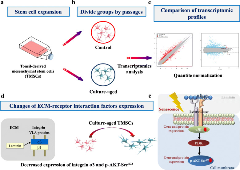
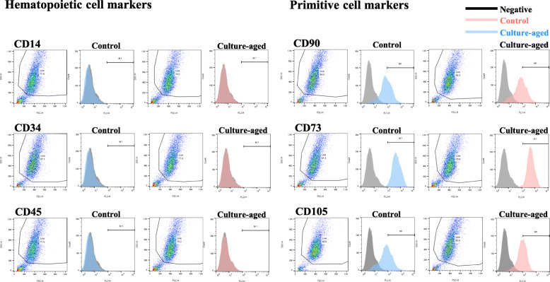
Tonsil-derived mesenchymal stem cells (TMSCs) with 20-25 passages were designated as culture-aged TMSCs, and their mesodermal differentiation potentials as well as markers of senescence and stemness were compared with the control TMSCs passaged up to 8 times at the most (designated as young). A whole-genome analysis was used to identify novel regulatory factors that distinguish between the culture-aged and control TMSCs. The identified markers of replicative senescence were validated using Western blot analyses.

RESULTS

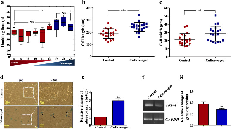
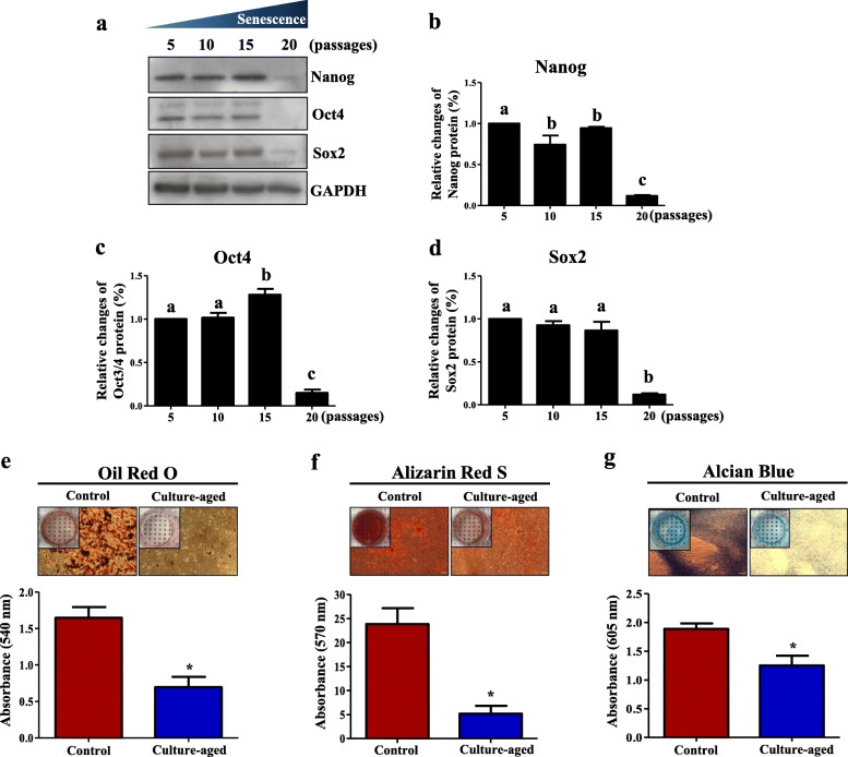
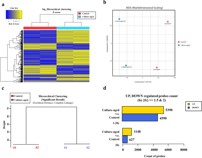
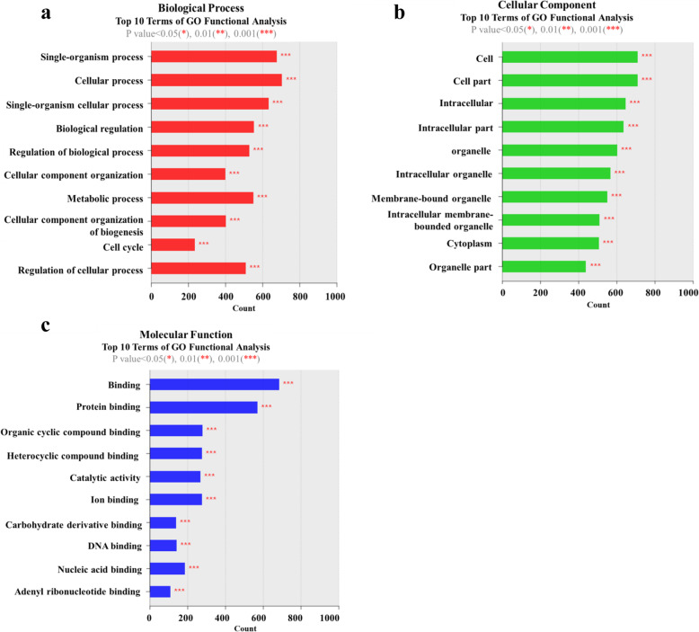
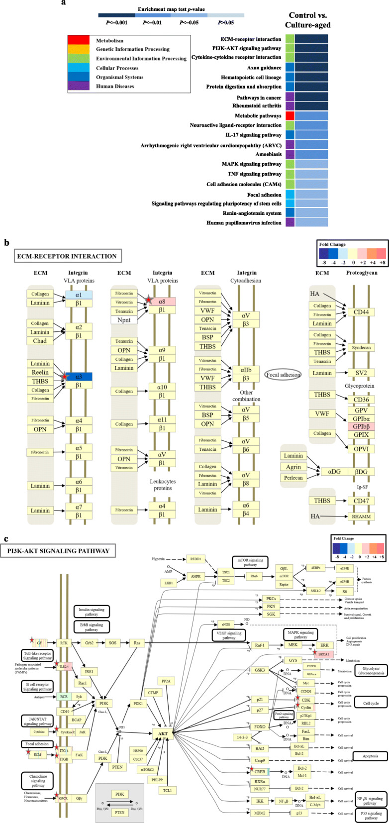
The culture-aged TMSCs showed longer doubling time compared to control TMSCs and had higher expression of senescence-associated (SA)-β-gal staining but lower expression of the stemness protein markers, including Nanog, Oct4, and Sox2 with decreased adipogenic, osteogenic, and chondrogenic differentiation potentials. Microarray analyses identified a total of 18,614 differentially expressed genes between the culture-aged and control TMSCs. The differentially expressed genes were classified into the Gene Ontology categories of cellular component (CC), functional component (FC), and biological process (BP) using KEGG (Kyoto encyclopedia of genes and genomes) pathway analysis. This analysis revealed that those genes associated with CC and BP showed the most significant difference between the culture-aged and control TMSCs. The genes related to extracellular matrix-receptor interactions were also shown to be significantly different (p < 0.001). We also found that culture-aged TMSCs had decreased expressions of integrin α3 (ITGA3) and phosphorylated AKT protein (p-AKT-Ser) compared to the control TMSCs.

CONCLUSIONS

Our data suggest that activation of ECM-receptor signaling, specifically involved with integrin family-mediated activation of the intracellular cell survival-signaling molecule AKT, can regulate stem cell senescence in TMSCs. Among these identified factors, ITGA3 was found to be a representative biomarker of the senescent TMSCs. Exclusion of the TMSCs with the senescent TMSC markers in this study could potentially increase the therapeutic efficacy of TMSCs in clinical applications.

摘要

背景

间充质干细胞(MSCs)已被广泛用于干细胞治疗,为了满足再生医学实际应用中对细胞数量的需求,通常需要对干细胞进行多次传代培养。长期的细胞传代培养可能会导致复制性衰老,从而导致干细胞功能和干性逐渐下降,失去多能性特征。为了提高干细胞治疗的疗效,确定衰老细胞的特异性标志物非常重要。

方法

将传代 20-25 次的扁桃体来源间充质干细胞(TMSCs)定义为培养老化的 TMSCs,与传代最多 8 次的对照组 TMSCs(定义为年轻组)相比,比较其中胚层分化潜能以及衰老和干性标志物。使用全基因组分析来鉴定区分培养老化和对照组 TMSCs 的新型调控因子。使用 Western blot 分析验证复制性衰老的鉴定标志物。

结果

与对照组 TMSCs 相比,培养老化的 TMSCs 的倍增时间更长,衰老相关-β-半乳糖染色(SA-β-gal)的表达更高,而干性蛋白标志物 Nanog、Oct4 和 Sox2 的表达水平较低,其成脂、成骨和软骨分化潜能降低。微阵列分析在培养老化和对照组 TMSCs 之间共鉴定出 18614 个差异表达基因。使用京都基因与基因组百科全书(KEGG)通路分析将差异表达基因分为细胞成分(CC)、功能成分(FC)和生物过程(BP)的基因本体论(GO)类别。该分析表明,那些与 CC 和 BP 相关的基因在培养老化和对照组 TMSCs 之间存在最显著的差异。与细胞外基质-受体相互作用相关的基因也表现出显著差异(p<0.001)。我们还发现,与对照组 TMSCs 相比,培养老化的 TMSCs 中整合素 α3(ITGA3)和磷酸化 AKT 蛋白(p-AKT-Ser)的表达降低。

结论

我们的数据表明,细胞外基质-受体信号的激活,特别是涉及整联蛋白家族介导的细胞内细胞存活信号分子 AKT 的激活,可以调节 TMSCs 的干细胞衰老。在这些鉴定出的因子中,ITGA3 被发现是衰老 TMSCs 的代表性标志物。在本研究中排除具有衰老 TMSC 标志物的 TMSCs 可能会增加 TMSCs 在临床应用中的治疗效果。

https://cdn.ncbi.nlm.nih.gov/pmc/blobs/d3f0/7430027/b4237753b52b/13287_2020_1860_Fig8_HTML.jpg
https://cdn.ncbi.nlm.nih.gov/pmc/blobs/d3f0/7430027/c9bcab312864/13287_2020_1860_Fig1_HTML.jpg
https://cdn.ncbi.nlm.nih.gov/pmc/blobs/d3f0/7430027/bd65147cb524/13287_2020_1860_Fig2_HTML.jpg
https://cdn.ncbi.nlm.nih.gov/pmc/blobs/d3f0/7430027/a7e36eed81fd/13287_2020_1860_Fig3_HTML.jpg
https://cdn.ncbi.nlm.nih.gov/pmc/blobs/d3f0/7430027/9f75eed19faa/13287_2020_1860_Fig4_HTML.jpg
https://cdn.ncbi.nlm.nih.gov/pmc/blobs/d3f0/7430027/02e892ca92b8/13287_2020_1860_Fig5_HTML.jpg
https://cdn.ncbi.nlm.nih.gov/pmc/blobs/d3f0/7430027/801e893a2ac1/13287_2020_1860_Fig6_HTML.jpg
https://cdn.ncbi.nlm.nih.gov/pmc/blobs/d3f0/7430027/28e408eeba50/13287_2020_1860_Fig7_HTML.jpg
https://cdn.ncbi.nlm.nih.gov/pmc/blobs/d3f0/7430027/b4237753b52b/13287_2020_1860_Fig8_HTML.jpg

相似文献

1
A transcriptomic analysis of serial-cultured, tonsil-derived mesenchymal stem cells reveals decreased integrin α3 protein as a potential biomarker of senescent cells.连续培养扁桃体来源间充质干细胞的转录组分析显示整合素 α3 蛋白减少,可作为衰老细胞的潜在生物标志物。
Stem Cell Res Ther. 2020 Aug 17;11(1):359. doi: 10.1186/s13287-020-01860-y.
2
Podoplanin depletion in tonsil-derived mesenchymal stem cells induces cellular senescence via regulation of the p16/Rb pathway.扁桃体来源的间充质干细胞中血小板源性生长因子受体β的缺失通过p16/Rb通路的调节诱导细胞衰老。
Cell Commun Signal. 2024 Jun 12;22(1):323. doi: 10.1186/s12964-024-01705-8.
3
Tensin-3 Regulates Integrin-Mediated Proliferation and Differentiation of Tonsil-Derived Mesenchymal Stem Cells.Tensin-3 通过整合素调节扁桃体来源间充质干细胞的增殖和分化。
Cells. 2019 Dec 30;9(1):89. doi: 10.3390/cells9010089.
4
Characterization of long-term in vitro culture-related alterations of human tonsil-derived mesenchymal stem cells: role for CCN1 in replicative senescence-associated increase in osteogenic differentiation.人扁桃体来源间充质干细胞长期体外培养相关改变的特征:CCN1在复制性衰老相关成骨分化增加中的作用
J Anat. 2014 Nov;225(5):510-8. doi: 10.1111/joa.12229. Epub 2014 Aug 23.
5
Proliferation-Related Features of the Human Mesenchymal Stem Cells Derived from Palatine Tonsils, Adipose Tissues, and Bone Marrow.人腭扁桃体、脂肪组织和骨髓间充质干细胞的增殖相关特征。
Tissue Eng Regen Med. 2023 Dec;20(7):1119-1132. doi: 10.1007/s13770-023-00564-7. Epub 2023 Aug 18.
6
Tonsil-derived mesenchymal stem cells incorporated in reactive oxygen species-releasing hydrogel promote bone formation by increasing the translocation of cell surface GRP78.扁桃体衍生间充质干细胞被包裹在具有释放活性氧功能的水凝胶中,通过增加细胞表面 GRP78 的易位来促进骨形成。
Biomaterials. 2021 Nov;278:121156. doi: 10.1016/j.biomaterials.2021.121156. Epub 2021 Sep 25.
7
Tonsil-derived mesenchymal stem cells enhance allogeneic bone marrow engraftment via collagen IV degradation.扁桃体来源间充质干细胞通过降解胶原 IV 增强同种异体骨髓植入。
Stem Cell Res Ther. 2021 Jun 5;12(1):329. doi: 10.1186/s13287-021-02414-6.
8
Therapeutic Effect of IL1β Priming Tonsil Derived-Mesenchymal Stem Cells in Osteoporosis.白介素 1β 预刺激扁桃体来源间充质干细胞对骨质疏松症的治疗作用。
Tissue Eng Regen Med. 2021 Oct;18(5):851-862. doi: 10.1007/s13770-021-00350-3. Epub 2021 Jun 11.
9
Matrix Metalloproteinase 1 as a Marker of Tonsil-Derived Mesenchymal Stem Cells to Assess Bone Marrow Cell Migration.基质金属蛋白酶 1 作为扁桃体衍生间充质干细胞的标志物评估骨髓细胞迁移。
Tissue Eng Regen Med. 2023 Apr;20(2):271-284. doi: 10.1007/s13770-022-00501-0. Epub 2022 Dec 3.
10
Human Tonsil-Derived Mesenchymal Stromal Cells Maintain Proliferating and ROS-Regulatory Properties via Stanniocalcin-1.人扁桃体间充质基质细胞通过 STC-1 维持增殖和 ROS 调节特性。
Cells. 2020 Mar 6;9(3):636. doi: 10.3390/cells9030636.

引用本文的文献

1
Development of Advanced Oral-on-a-Chip: Replicating the Intricate Human Oral Microenvironment.先进口腔芯片的开发:复制复杂的人类口腔微环境。
Int J Biol Sci. 2024 Oct 28;20(15):5888-5909. doi: 10.7150/ijbs.104351. eCollection 2024.
2
Integrin αVβ3 antagonist-c(RGDyk) peptide attenuates the progression of ossification of the posterior longitudinal ligament by inhibiting osteogenesis and angiogenesis.整合素 αVβ3 拮抗剂-c(RGDyk) 肽通过抑制成骨和血管生成来减轻后纵韧带骨化的进展。
Mol Med. 2024 May 2;30(1):57. doi: 10.1186/s10020-024-00822-x.
3
Ethanol induces replication fork stalling and membrane stress in immortalized laryngeal cells.

本文引用的文献

1
Optimization of Microenvironments Inducing Differentiation of Tonsil-Derived Mesenchymal Stem Cells into Endothelial Cell-Like Cells.优化微环境诱导扁桃体来源间充质干细胞向内皮细胞样细胞分化。
Tissue Eng Regen Med. 2019 Oct 30;16(6):631-643. doi: 10.1007/s13770-019-00221-y. eCollection 2019 Dec.
2
Integrin-α3 Is a Functional Marker of Ex Vivo Expanded Human Long-Term Hematopoietic Stem Cells.整合素-α3 是体外扩增的人长期造血干/祖细胞的功能标志物。
Cell Rep. 2019 Jul 23;28(4):1063-1073.e5. doi: 10.1016/j.celrep.2019.06.084.
3
Application of Tonsil-Derived Mesenchymal Stem Cells in Tissue Regeneration: Concise Review.
乙醇在永生化喉细胞中诱导复制叉停滞和膜应激。
iScience. 2023 Nov 23;26(12):108564. doi: 10.1016/j.isci.2023.108564. eCollection 2023 Dec 15.
4
Efficient expansion and delayed senescence of hUC-MSCs by microcarrier-bioreactor system.微载体-生物反应器系统高效扩增及延缓 hUC-MSCs 衰老。
Stem Cell Res Ther. 2023 Oct 4;14(1):284. doi: 10.1186/s13287-023-03514-1.
5
Cyclin-Dependent Kinase 1 Inhibition Potentiates the Proliferation of Tonsil-Derived Mesenchymal Stem Cells by Delaying Cellular Senescence.细胞周期蛋白依赖性激酶1抑制通过延迟细胞衰老增强扁桃体来源间充质干细胞的增殖。
Stem Cells Int. 2022 Jul 21;2022:4302992. doi: 10.1155/2022/4302992. eCollection 2022.
6
BMP9 reduces age-related bone loss in mice by inhibiting osteoblast senescence through Smad1-Stat1-P21 axis.骨形态发生蛋白9通过Smad1-Stat1-P21轴抑制成骨细胞衰老,从而减少小鼠与年龄相关的骨质流失。
Cell Death Discov. 2022 May 6;8(1):254. doi: 10.1038/s41420-022-01048-8.
7
Cellular senescence in lymphoid organs and immunosenescence.淋巴器官中的细胞衰老与免疫衰老。
Aging (Albany NY). 2021 Aug 12;13(15):19920-19941. doi: 10.18632/aging.203405.
8
Engineering osteoarthritic cartilage model through differentiating senescent human mesenchymal stem cells for testing disease-modifying drugs.通过分化衰老的人骨髓间充质干细胞来构建骨关节炎软骨模型,用于测试疾病修饰药物。
Sci China Life Sci. 2022 Feb;65(2):309-327. doi: 10.1007/s11427-021-1933-7. Epub 2021 Jun 4.
扁桃体来源间充质干细胞在组织再生中的应用:简要综述。
Stem Cells. 2019 Oct;37(10):1252-1260. doi: 10.1002/stem.3058. Epub 2019 Jul 25.
4
Differentiation of Motor Neuron-Like Cells from Tonsil-Derived Mesenchymal Stem Cells and Their Possible Application to Neuromuscular Junction Formation.扁桃体来源间充质干细胞向运动神经元样细胞的分化及其在神经肌肉接头形成中的可能应用。
Int J Mol Sci. 2019 Jun 1;20(11):2702. doi: 10.3390/ijms20112702.
5
Methods and Strategies for Procurement, Isolation, Characterization, and Assessment of Senescence of Human Mesenchymal Stem Cells from Adipose Tissue.从脂肪组织中获取、分离、鉴定和评估人间充质干细胞衰老的方法与策略
Methods Mol Biol. 2019;2045:37-92. doi: 10.1007/7651_2018_174.
6
Conditioned Medium from Tonsil-Derived Mesenchymal Stem Cells Relieves CCl-Induced Liver Fibrosis in Mice.扁桃体来源的间充质干细胞条件培养基可缓解小鼠四氯化碳诱导的肝纤维化
Tissue Eng Regen Med. 2018 Oct 19;16(1):51-58. doi: 10.1007/s13770-018-0160-8. eCollection 2019 Feb.
7
Tissue Regeneration of Human Mesenchymal Stem Cells on Porous Gelatin Micro-Carriers by Long-Term Dynamic Culture.多孔明胶微载体长期动态培养对人骨髓间充质干细胞的组织再生。
Tissue Eng Regen Med. 2019 Jan 28;16(1):19-28. doi: 10.1007/s13770-018-00174-8. eCollection 2019 Feb.
8
Expression of tenocyte lineage-related factors from tonsil-derived mesenchymal stem cells.扁桃体来源的间充质干细胞中肌腱细胞谱系相关因子的表达
Tissue Eng Regen Med. 2016 Apr 5;13(2):162-170. doi: 10.1007/s13770-016-9134-x. eCollection 2016 Apr.
9
Signaling of extracellular matrices for tissue regeneration and therapeutics.用于组织再生和治疗的细胞外基质信号传导。
Tissue Eng Regen Med. 2016 Feb 2;13(1):1-12. doi: 10.1007/s13770-016-9075-0. eCollection 2016 Feb.
10
Mesenchymal Stromal/Stem Cells in Regenerative Medicine and Tissue Engineering.再生医学与组织工程中的间充质基质/干细胞
Stem Cells Int. 2018 Aug 19;2018:8031718. doi: 10.1155/2018/8031718. eCollection 2018.