Department of Nuclear Medicine and PET-Centre, Aarhus University Hospital, Aarhus, Denmark.
Department of Clinical Medicine, Aarhus University, Aarhus, Denmark.
Eur J Nucl Med Mol Imaging. 2021 Feb;48(2):532-542. doi: 10.1007/s00259-020-04998-2. Epub 2020 Aug 18.
Tumour blood flow (TBF) is a crucial determinant of cancer growth. Recently, we validated Rubidium-82 (Rb) positron emission tomography (PET) for TBF measurement in prostate cancer (PCa) and found TBF and cancer aggressiveness positively correlated. The aims of the present study were to determine the ability of TBF for separating significant from insignificant PCa and to examine the relation to underlying Na/K-ATPase density, which is relevant as Rb is transported intracellularly via the Na/K-ATPase.
One hundred and two patients were included for pelvic Rb PET scan prior to magnetic resonance imaging (MRI)-guided prostate biopsy. Findings constituted 100 PCa lesions (86 patients) and 25 benign lesions (16 patients). Tumours were defined on MRI and transferred to Rb PET for TBF measurement. Immunohistochemical Na/K-ATPase staining was subsequently performed on biopsies.
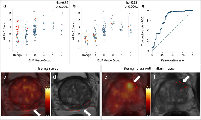
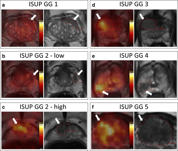
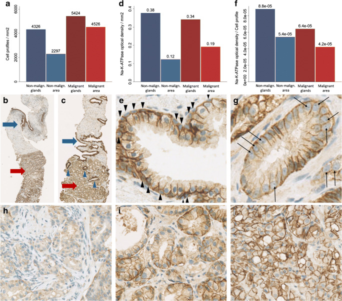
TBF was the superior predictor (rho = 0.68, p < 0.0001, inflammatory lesions excluded) of MRI-guided biopsy grade group (GG) over lowest apparent diffusion coefficient (ADC) value (rho = -0.23, p = 0.01), independent of ADC value and tumour volume (p < 0.0001). PET could separate GG-2-5 from GG-1 and benign lesions with an area under the curve (AUC), sensitivity, and specificity of 0.79, 96%, and 59%, respectively. For separating GG-3-5 from GG-1-2 and benign lesions the AUC, sensitivity, and specificity were 0.82, 95%, and 63%, respectively. Na/K-ATPase density per PCa cell profile was 38% lower compared with that of the benign prostate cell profiles. Neither cell density nor Na/K-ATPase density determined tumour Rb uptake.
TBF is an independent predictor of PCa aggressiveness and deserves more attention, as it may be valuable in separating clinically significant from insignificant PCa.
肿瘤血流(TBF)是癌症生长的关键决定因素。最近,我们验证了钌-82(Rb)正电子发射断层扫描(PET)在前列腺癌(PCa)中的 TBF 测量,并发现 TBF 与癌症侵袭性呈正相关。本研究的目的是确定 TBF 分离有意义和无意义 PCa 的能力,并检查与潜在的 Na/K-ATPase 密度的关系,因为 Rb 通过 Na/K-ATPase 在内质网内运输。
102 名患者接受盆腔 Rb PET 扫描,然后进行磁共振成像(MRI)引导的前列腺活检。结果包括 100 例 PCa 病变(86 例患者)和 25 例良性病变(16 例患者)。在 MRI 上定义肿瘤,并将其转移到 Rb PET 进行 TBF 测量。随后对活检进行 Na/K-ATPase 免疫组织化学染色。
TBF 是 MRI 引导活检分级组(GG)的优越预测因子(rho=0.68,p<0.0001,排除炎症病变),优于最低表观扩散系数(ADC)值(rho=-0.23,p=0.01),独立于 ADC 值和肿瘤体积(p<0.0001)。PET 可以将 GG-2-5 与 GG-1 和良性病变分开,曲线下面积(AUC)、敏感性和特异性分别为 0.79、96%和 59%。将 GG-3-5 与 GG-1-2 和良性病变分开时,AUC、敏感性和特异性分别为 0.82、95%和 63%。每个 PCa 细胞的 Na/K-ATPase 密度比良性前列腺细胞的密度低 38%。细胞密度和 Na/K-ATPase 密度均不能决定肿瘤摄取 Rb。
TBF 是 PCa 侵袭性的独立预测因子,值得更多关注,因为它可能有助于区分有临床意义和无意义的 PCa。