Deng Minghong, Lin Chunwang, Zeng Xianglin, Zhang Jianping, Wen Fang, Liu Ziguang, Wu Haiyan, Wu Xiaofeng
Pediatric Intensive Care Unit, Shunde Women and Children's Hospital of Guangdong Medical University, Foshan, Guangdong, China (mainland).
Pathological Department, Shunde Women and Children's Hospital of Guangdong Medical University, Foshan, Guangdong, China (mainland).
Med Sci Monit. 2020 Aug 21;26:e922429. doi: 10.12659/MSM.922429.
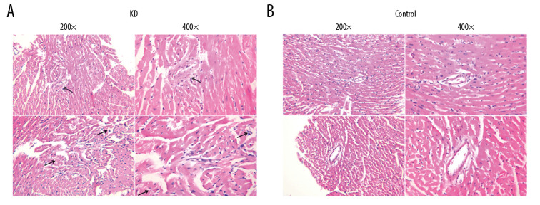
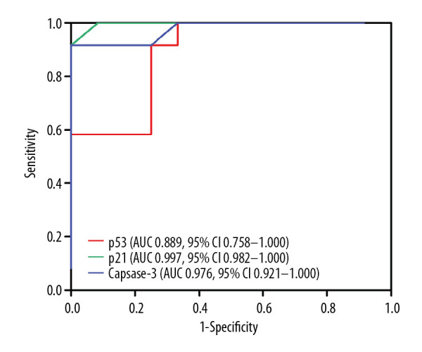
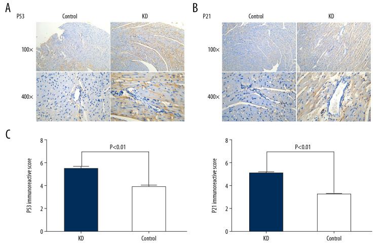
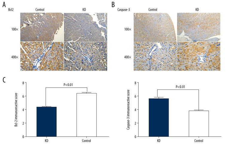
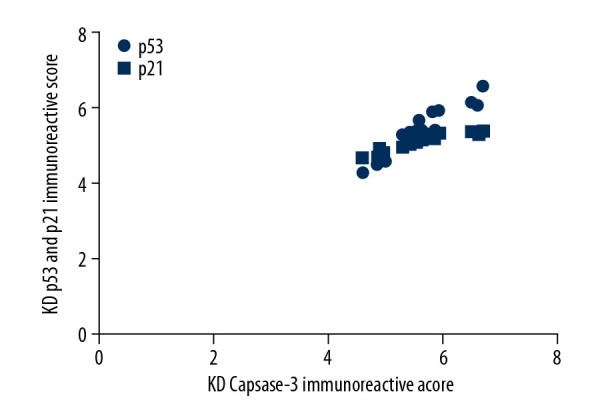
BACKGROUND Overexpression of p53, p21, and caspase-3 promotes apoptosis of vascular smooth muscle cells. However, the mechanisms that lead to apoptosis of coronary artery smooth muscle cells (CASMCs) is unclear in Kawasaki disease (KD). This study investigated involvement of p53, p21, and caspase-3 in the apoptosis of CASMCs from a Kawasaki vasculitis mouse model. MATERIAL AND METHODS The Kawasaki vasculitis mouse model with coronary artery lesions was generated via administration of Lactobacillus casei cell wall extract. In 2 groups of mice (healthy control and KD vasculitis mice), the levels of p53, p21, and caspase-3 protein in the root of the coronary artery were evaluated via immunohistochemistry. Receiver operating characteristic curves were plotted for determination of area under the curve, 95% confidence interval, sensitivity, specificity, and cutoff values for the ability of p53, p21, and caspase-3 expression to predict CASMC apoptosis and coronary artery lesion formation in KD vasculitis mice. RESULTS Compared with healthy mice, KD vasculitis mice had a significantly higher apoptosis index and upregulated p53, p21, and caspase-3 expression. Also, the immunoreactive score for caspase-3 was positively correlated with the immunoreactivity scores for p53 and p21. The optimal cutoff values for p53, p21, and caspase-3 expression for predicting the presence of coronary artery lesions were 4.15, 4.18, and 4.22, respectively. CONCLUSIONS Upregulated levels of p53, p21, and caspase-3 promoted apoptosis of CASMCs in KD vasculitis mice. Thus, the levels of p53, p21, and caspase-3 may serve as valuable predictors of coronary artery lesion formation in KD.
p53、p21和半胱天冬酶-3的过表达促进血管平滑肌细胞凋亡。然而,川崎病(KD)中导致冠状动脉平滑肌细胞(CASMCs)凋亡的机制尚不清楚。本研究调查了p53、p21和半胱天冬酶-3在川崎病血管炎小鼠模型的CASMCs凋亡中的作用。
通过给予干酪乳杆菌细胞壁提取物建立具有冠状动脉病变的川崎病血管炎小鼠模型。在两组小鼠(健康对照和KD血管炎小鼠)中,通过免疫组织化学评估冠状动脉根部p53、p21和半胱天冬酶-3蛋白的水平。绘制受试者工作特征曲线,以确定曲线下面积、95%置信区间、敏感性、特异性以及p53、p21和半胱天冬酶-3表达预测KD血管炎小鼠CASMC凋亡和冠状动脉病变形成能力的截断值。
与健康小鼠相比,KD血管炎小鼠的凋亡指数显著更高,且p53、p21和半胱天冬酶-3表达上调。此外,半胱天冬酶-3的免疫反应评分与p53和p21的免疫反应评分呈正相关。预测冠状动脉病变存在的p53、p21和半胱天冬酶-3表达的最佳截断值分别为4.15、4.18和4.22。
p53、p21和半胱天冬酶-3水平上调促进了KD血管炎小鼠CASMCs的凋亡。因此,p53、p21和半胱天冬酶-3的水平可能是KD中冠状动脉病变形成的有价值预测指标。