Shamurailatpam Dayananda Sharma, Manikandan A, Ganapathy K, Noufal M P, Patro Kartikeshwar C, Rajesh T, Jalali R
Department of Medical Physics, Apollo Proton Cancer Centre, Chennai, Tamil Nadu, India.
Department of Radiation Oncology, Apollo Proton Cancer Centre, Chennai, Tamil Nadu, India.
J Med Phys. 2020 Apr-Jun;45(2):59-65. doi: 10.4103/jmp.JMP_12_20. Epub 2020 Jul 20.
The purpose of this study is to evaluate the performance characteristic of volumetric image-guided dedicated-nozzle pencil beam-scanning proton therapy (PT) system.
PT system was characterized for electromechanical, image quality, and registration accuracy. Proton beam of 70.2-226.2 MeV was characterized for short- and long-term reproducibility in integrated depth dose; spot profile characteristics at different air gap and gantry angle; positioning accuracy of single and pattern of spot; dose linearity, reproducibility and consistency. All measurements were carried out using various X-ray and proton-beam specific detectors following standard protocols.
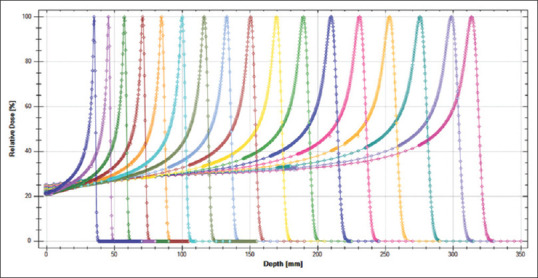
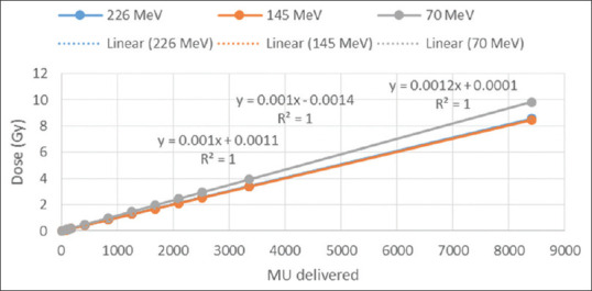
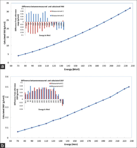
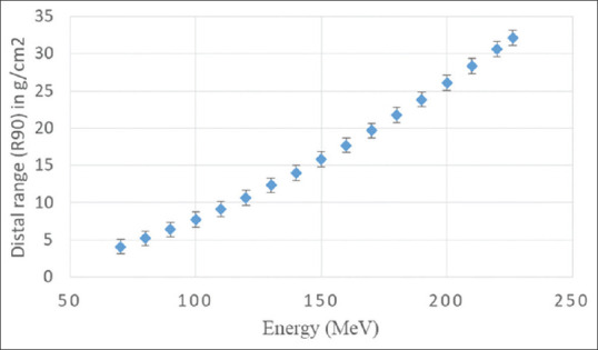
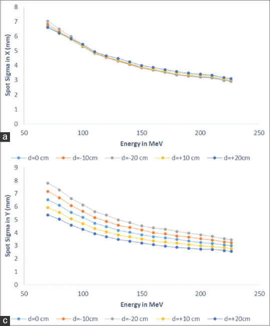
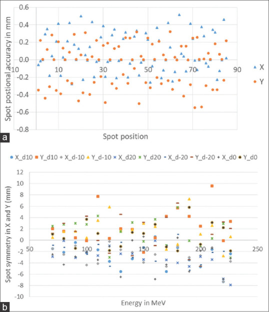
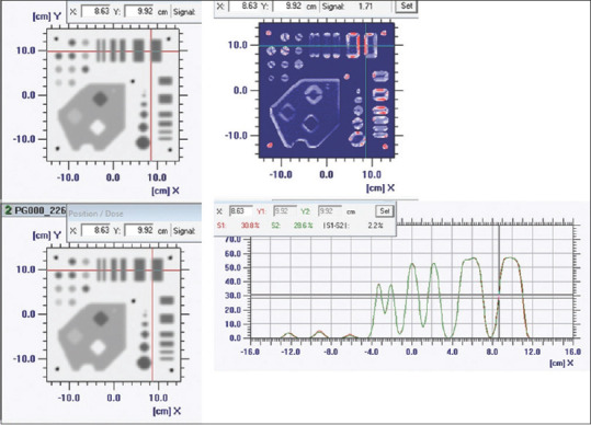
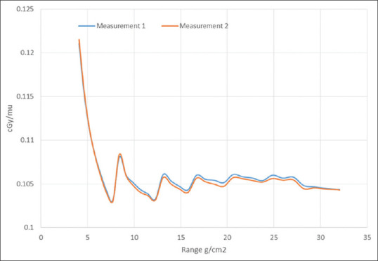
All electro-mechanical, imaging, and safety parameters performed well within the specified tolerance limit. The image registration errors along three translation and three rotational axes were ≤0.5 mm and ≤0.2° for both point-based and intensity-based auto-registration. Distal range (R) and distal dose fall-off (DDF) of 70.2-226.2 MeV proton beams were within 1 mm of calculated values based on the international commission on radiation units and measurements 49 and 0.0156× R, respectively. The R and DDF were reproducible within a standard deviation of 0.05 g/cm during the first 8 months. Dose were linear from 18.5 (0.011 MU/spot) to 8405 (5 MU/spot) MU, reproducible within 0.5% in 5 consecutive days and consistent within 0.8% for full rotation. The cGy/MU for 70.2-226.2MeV was consistent within 0.5%. In-air X(Y) spot-sigma at isocenter varies from 2.96 (3.00) mm to 6.68 (6.52) mm for 70.2-226.2 MeV. Maximum variation of spot-sigma with air-gap of ±20 cm was ±0.36 mm (5.28%) and ±0.82 mm (±12.5%) along X- and Y-direction and 3.56% for full rotation. Relative spot positions were accurate within ±0.6 mm. The planned and delivered spot pattern of known complex geometry agreed with (γ%≤1) for 1% @ 1 mm >98% for representative five-proton energies at four gantry angle.
The PT-system performed well within the expected accuracy level and consistent over a period of 8 months. The methodology and data presented here may help upcoming modern PT center during their crucial phase of commissioning.
本研究旨在评估容积图像引导专用喷嘴笔形束扫描质子治疗(PT)系统的性能特征。
对PT系统的机电性能、图像质量和配准精度进行了表征。对能量为70.2 - 226.2 MeV的质子束在累积深度剂量方面的短期和长期可重复性、不同气隙和机架角度下的束斑轮廓特征、单个束斑及束斑图案的定位精度、剂量线性、可重复性和一致性进行了表征。所有测量均按照标准方案使用各种X射线和质子束专用探测器进行。
所有机电、成像和安全参数均在规定的公差范围内表现良好。基于点和基于强度的自动配准在三个平移轴和三个旋转轴上的图像配准误差均≤0.5 mm且≤0.2°。70.2 - 226.2 MeV质子束的远端射程(R)和远端剂量下降(DDF)分别在基于国际辐射单位与测量委员会第49号报告计算值的1 mm范围内和0.0156×R以内。在最初8个月内,R和DDF的可重复性标准差为0.05 g/cm。剂量在18.5(0.011 MU/束斑)至8405(5 MU/束斑)MU范围内呈线性,在连续5天内的可重复性为0.5%以内,在全旋转范围内的一致性为0.8%以内。70.2 - 226.2 MeV的cGy/MU一致性在0.5%以内。对于70.2 - 226.2 MeV,等中心处空气中X(Y)方向的束斑标准差从2.96(3.00)mm变化到6.68(6.52)mm。束斑标准差随±20 cm气隙在X和Y方向上的最大变化分别为±0.36 mm(5.28%)和±0.82 mm(±12.5%),全旋转时为3.56%。相对束斑位置的精度在±0.6 mm以内。对于四个机架角度下的代表性五种质子能量而言,已知复杂几何形状的计划和交付束斑图案符合(γ%≤1),即1%@1 mm>98%。
PT系统在预期精度水平内表现良好,且在8个月的时间内保持一致。本文介绍的方法和数据可能有助于即将建成的现代PT中心在其关键的调试阶段。