Ophthalmology Clinic, Department of Medicine and Science of Ageing, University G. D'Annunzio Chieti-Pescara, Chieti, via dei Vestini 31, 66100, Italy.
Department of Neuroscience, Imaging, and Clinical Sciences, University G. D'Annunzio Chieti-Pescara, Chieti, via dei Vestini 31, 66100, Italy.
Transl Vis Sci Technol. 2020 Jun 11;9(7):13. doi: 10.1167/tvst.9.7.13. eCollection 2020 Jun.
To evaluate the changes of retinal capillary nonperfusion areas and retinal capillary vessel density of the superficial capillary plexus (SCP) and deep capillary plexus in patients with diabetes with diabetic macular edema treated with an intravitreal dexamethasone implant (IDI).
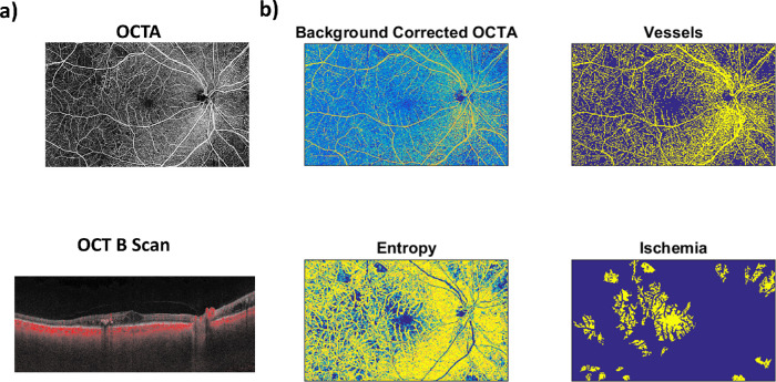
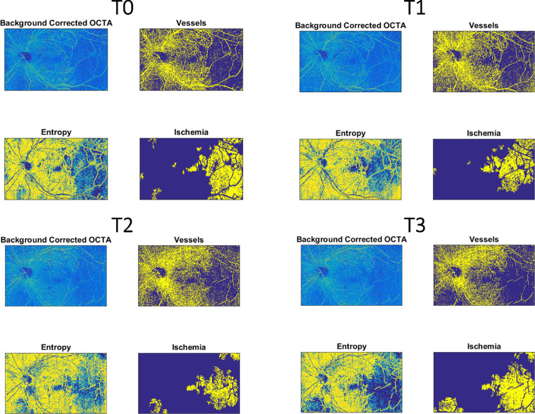
We enrolled 28 patients with diabetic retinopathy and diabetic macular edema candidates to IDI. All patients underwent widefield optical coherence tomography angiography with PLEX Elite 9000 device with 15 × 9 mm scans centered on the foveal center at baseline, 1 month, 2 months, and 4 months after IDI. In all the patients, the variation of the retinal capillary nonperfusion areas and of the retinal vessel density of the SCP and deep capillary plexus were calculated using an automatic software written in Matlab (MathWorks, Natick, MA).
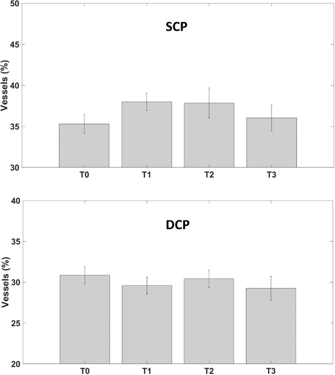
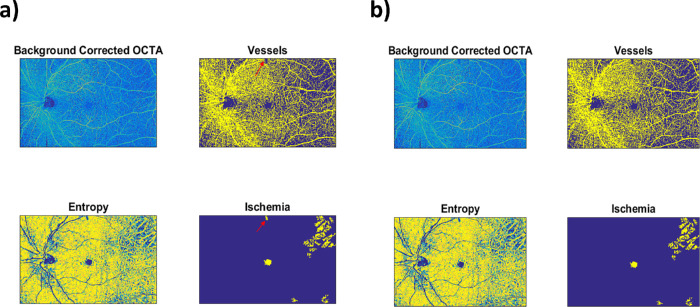
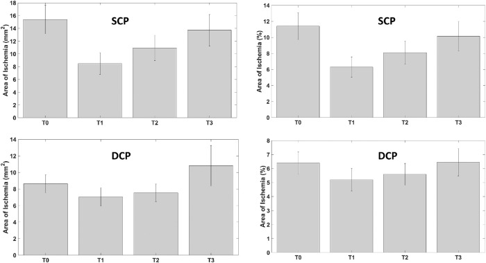
During follow-up, SCP showed a statistically significant reduction of ischemic areas at 1 month after IDI ( = 0.04) and slightly increased not significantly thereafter ( = 0.15). The percentage of nonperfusion areas changed from 11.4% at baseline, to 6.3% at 1 month, 8.1%, at 2 months, and 10.2% at 4 months. The whole vessel density of SCP slightly increased (not significantly) from 35.30% at baseline to 38.00% at 1 month, and then decreased to 37.85% at 2 months and 36.04% at 4 months ( = 0.29). Retinal capillary nonperfusion areas and retinal vessel density at the deep capillary plexus did not change significantly ( = 0.31 and = 0.73, respectively).
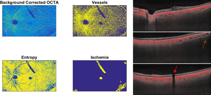
Widefield optical coherence tomography angiography showed a decrease in retinal capillary nonperfusion areas after dexamethasone implant suggesting a possible drug-related reperfusion of retinal capillaries particularly evident in the early period.
A custom-made automatic analysis of retinal nonperfusion areas may allow a better and precise evaluation of ischemic changes after intravitreal therapy.
评估玻璃体内注射地塞米松植入物(IDI)治疗糖尿病黄斑水肿患者后视网膜毛细血管无灌注区和浅层毛细血管丛(SCP)及深层毛细血管丛视网膜毛细血管密度的变化。
我们招募了 28 名糖尿病性视网膜病变和糖尿病黄斑水肿候选患者进行 IDI。所有患者均在基线、IDI 后 1 个月、2 个月和 4 个月时使用 PLEX Elite 9000 设备进行广角光相干断层扫描血管造影,以黄斑中心为中心进行 15×9mm 扫描。在所有患者中,使用 Matlab(马萨诸塞州纳蒂克的 MathWorks)编写的自动软件计算视网膜毛细血管无灌注区和 SCP 及深层毛细血管丛视网膜血管密度的变化。
在随访期间,IDI 后 1 个月 SCP 出现缺血区的统计学显著减少( = 0.04),此后略有增加但无统计学意义( = 0.15)。无灌注区百分比从基线时的 11.4%变化至 1 个月时的 6.3%,2 个月时的 8.1%和 4 个月时的 10.2%。SCP 的全血管密度从基线时的 35.30%略有增加(无统计学意义)至 1 个月时的 38.00%,然后在 2 个月时降至 37.85%,在 4 个月时降至 36.04%( = 0.29)。深层毛细血管丛的视网膜毛细血管无灌注区和视网膜血管密度无明显变化( = 0.31 和 = 0.73)。
广角光相干断层扫描血管造影显示,IDI 后视网膜毛细血管无灌注区减少,提示可能与药物相关的视网膜毛细血管再灌注,尤其是在早期更为明显。
张宇轩