Laboratory of Immunopharmacology, Oswaldo Cruz Institute, Oswaldo Cruz Foundation, Fiocruz, Av. Brasil, 4365, Pavilhão 108, sala 45, Manguinhos, Rio de Janeiro, RJ, 21040-360, Brazil.
Laboratory of Pulmonary Investigation, Carlos Chagas Filho Institute of Biophysics, Federal University of Rio de Janeiro, Rio de Janeiro, Brazil.
Stem Cell Res Ther. 2020 Aug 26;11(1):367. doi: 10.1186/s13287-020-01874-6.
Malaria is one of the most critical global infectious diseases. Severe systemic inflammatory diseases, such as cerebral malaria, lead to the development of cognitive and behavioral alterations, such as learning disabilities and loss of memory capacity, as well as increased anxiety and depression. The consequences are profound and usually contribute to reduce the patient's quality of life. There are no therapies to treat the neurological sequelae of cerebral malaria. Mesenchymal stromal cells (MSCs) may be an alternative, since they have been used as therapy for neurodegenerative diseases and traumatic lesions of the central nervous system. So far, no study has investigated the effects of MSC therapy on the blood-brain barrier, leukocyte rolling and adherence in the brain, and depression like-behavior in experimental cerebral malaria.
Male C57BL/6 mice were infected with Plasmodium berghei ANKA (PbA, 1 × 10 PbA-parasitized red blood cells, intraperitoneally). At day 6, PbA-infected animals received chloroquine (25 mg/kg orally for seven consecutive days) as the antimalarial treatment and were then randomized to receive MSCs (1 × 10 cells in 0.05 ml of saline/mouse) or saline (0.05 ml) intravenously. Parasitemia, clinical score, and survival rate were analyzed throughout the experiments. Evans blue assay was performed at 6, 7, and 15 days post-infection (dpi). Behavioral tests were performed at 5 and 15 dpi. Intravital microscopy experiments and brain-derived neurotrophic factor (BDNF) protein expression analyses were performed at 7 dpi, whereas inflammatory mediators were measured at 15 dpi. In vitro, endothelial cells were used to evaluate the effects of conditioned media derived from MSCs (CMMSC) on cell viability by lactate dehydrogenase (LDH) release.
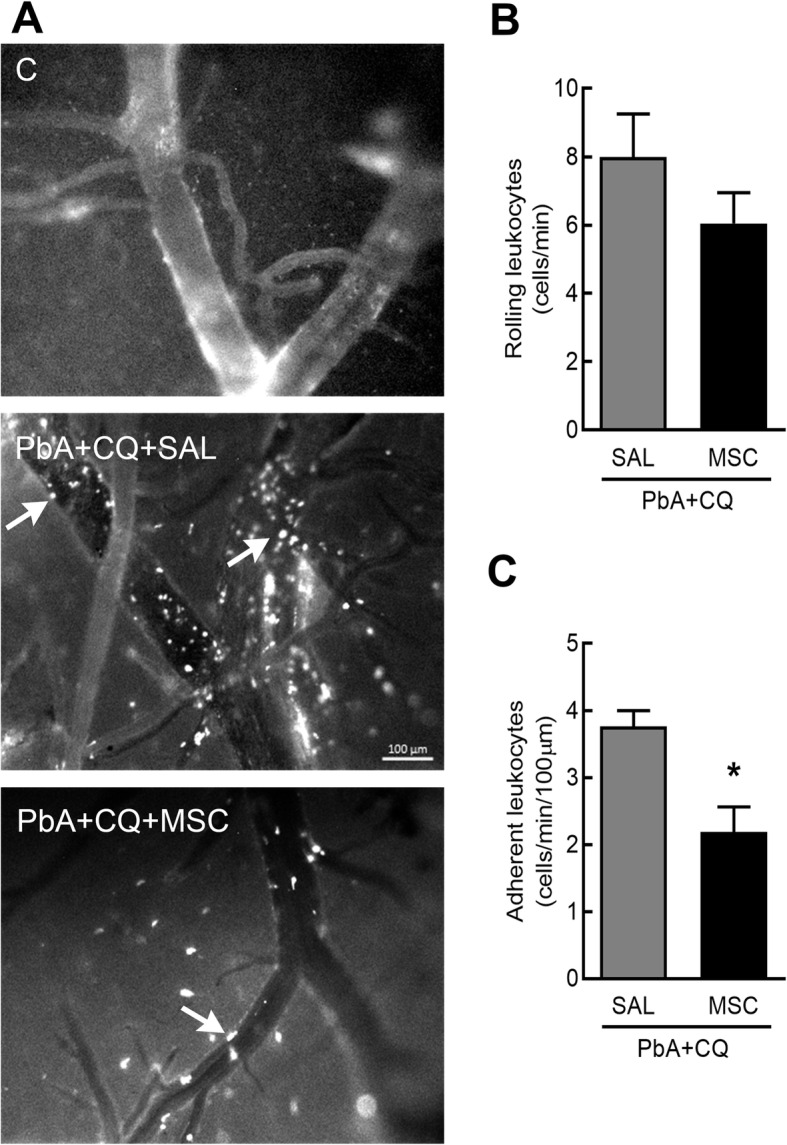
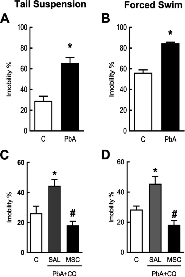
PbA-infected mice presented increased parasitemia, adherent leukocytes, blood-brain barrier permeability, and reduced BDNF protein levels, as well as depression-like behavior. MSCs mitigated behavioral alterations, restored BDNF and transforming growth factor (TGF)-β protein levels, and reduced blood-brain barrier dysfunction and leukocyte adhesion in the brain microvasculature. In a cultured endothelial cell line stimulated with heme, CMMSC reduced LDH release, suggesting a paracrine mechanism of action.
A single dose of MSCs as adjuvant therapy protected against vascular damage and improved depression-like behavior in mice that survived experimental cerebral malaria.
疟疾是最严重的全球性传染病之一。严重的全身性炎症性疾病,如脑疟疾,会导致认知和行为改变,如学习障碍和记忆能力丧失,以及焦虑和抑郁增加。其后果是深远的,通常会降低患者的生活质量。目前尚无治疗脑疟疾神经后遗症的方法。间充质基质细胞(MSCs)可能是一种替代方法,因为它们已被用于治疗神经退行性疾病和中枢神经系统创伤性损伤。到目前为止,还没有研究调查 MSC 治疗对实验性脑疟疾血脑屏障、白细胞滚动和黏附、抑郁样行为的影响。
雄性 C57BL/6 小鼠感染伯氏疟原虫 ANKA(PbA,1×10 个疟原虫感染的红细胞,腹腔内)。在第 6 天,PbA 感染的动物接受氯喹(连续 7 天口服 25mg/kg)作为抗疟治疗,然后随机接受 MSCs(1×10 个细胞,0.05ml 生理盐水/只)或生理盐水(0.05ml)静脉注射。整个实验过程中分析寄生虫血症、临床评分和存活率。在感染后 6、7 和 15 天进行 Evans 蓝测定。在 5 和 15 天进行行为测试。在 7 天进行活体显微镜实验和脑源性神经营养因子(BDNF)蛋白表达分析,而在 15 天进行炎症介质测量。在体外,使用内皮细胞评估 MSC 来源的条件培养基(CMMSC)对细胞活力的影响,通过乳酸脱氢酶(LDH)释放来评估。
PbA 感染的小鼠出现寄生虫血症增加、黏附白细胞、血脑屏障通透性增加、BDNF 蛋白水平降低以及抑郁样行为。MSCs 减轻了行为改变,恢复了 BDNF 和转化生长因子(TGF)-β蛋白水平,并减少了脑微血管中的血脑屏障功能障碍和白细胞黏附。在被血红素刺激的培养内皮细胞系中,CMMSC 减少了 LDH 的释放,表明存在旁分泌作用机制。
单次 MSC 剂量作为辅助治疗可防止血管损伤,并改善实验性脑疟疾中存活小鼠的抑郁样行为。