Department of Radiation Oncology, the First Affiliated Hospital of Soochow University, Suzhou, China.
Department of Oncology, the First Affiliated Hospital of Soochow University, Suzhou, China.
BMC Cancer. 2020 Aug 26;20(1):812. doi: 10.1186/s12885-020-07326-x.
Microwave ablation (MWA) is widely used to treat unresectable primary and secondary malignancies of the liver, and a limited number of studies indicate that ablation can cause not only necrosis at the in situ site but also an immunoreaction of the whole body. This study aimed to investigate the effects of MWA on cytokines in patients who underwent MWA for a hepatic malignancy.
Patients admitted to the Oncology Department in the First Affiliated Hospital of Soochow University between June 2015 and February 2019 were selected. Peripheral blood was collected from patients with a hepatic malignancy treated with MWA. The levels of cytokines (IL-2, IFN-γ, TNF-α, IL-12 p40, IL-12 p70, IL-4, IL-6, IL-8, IL-10, and vascular endothelial growth factor (VEGF)) were detected with a Milliplex® MAP Kit. The comparison times were as follows: before ablation, 24 h after ablation, 15 days after ablation, and 30 days after ablation. Data were analyzed using a paired sample t-tests and Spearman's correlation analysis.
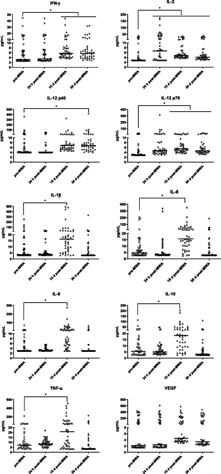
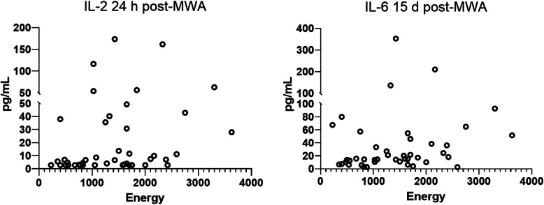
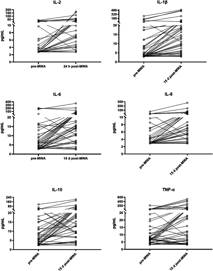
A total of 43 patients with hepatic malignancies were assessed. There were significant differences in IL-2, IL-12 p40, IL-12 p70, IL-1β, IL-8, and TNF-α at 24 h after MWA. Significant increases (> 2-fold vs. before ablation) were observed in IL-2, IL-1β, IL-6, IL-8, IL-10, and TNF-α after MWA. Elevated IL-2 and IL-6 levels after ablation were positively correlated with energy output during the MWA procedure.
WA treatment for hepatic malignancies can alter the serum levels of several cytokines such as IL-2 and IL-6.
微波消融(MWA)广泛用于治疗不可切除的原发性和继发性肝恶性肿瘤,少数研究表明消融不仅能引起原位组织坏死,还能引起全身免疫反应。本研究旨在探讨 MWA 对接受肝恶性肿瘤 MWA 治疗的患者细胞因子的影响。
选取 2015 年 6 月至 2019 年 2 月苏州大学第一附属医院肿瘤科收治的肝恶性肿瘤患者。采集 MWA 治疗的肝恶性肿瘤患者外周血,采用 Milliplex®MAP 试剂盒检测细胞因子(IL-2、IFN-γ、TNF-α、IL-12 p40、IL-12 p70、IL-4、IL-6、IL-8、IL-10 和血管内皮生长因子(VEGF))水平。比较时间点分别为消融前、消融后 24 h、消融后 15 d 和消融后 30 d。采用配对样本 t 检验和 Spearman 相关分析进行数据分析。
共评估了 43 例肝恶性肿瘤患者。MWA 后 24 h 时,IL-2、IL-12 p40、IL-12 p70、IL-1β、IL-8 和 TNF-α 差异有统计学意义。MWA 后,IL-2、IL-1β、IL-6、IL-8、IL-10 和 TNF-α 显著升高(与消融前相比,增加>2 倍)。消融后升高的 IL-2 和 IL-6 水平与 MWA 过程中的能量输出呈正相关。
WA 治疗肝恶性肿瘤可改变 IL-2 和 IL-6 等几种细胞因子的血清水平。