Laboratory of Bacterial Polysaccharides, Division of Bacterial Parasitic and Allergenic Products, U.S. Food and Drug Administration, Silver Spring, MD, United States.
Microscopy and Imaging Core Facility, Center for Biologics Evaluation and Research, U.S. Food and Drug Administration, Silver Spring, MD, United States.
Front Immunol. 2020 Jul 28;11:1569. doi: 10.3389/fimmu.2020.01569. eCollection 2020.
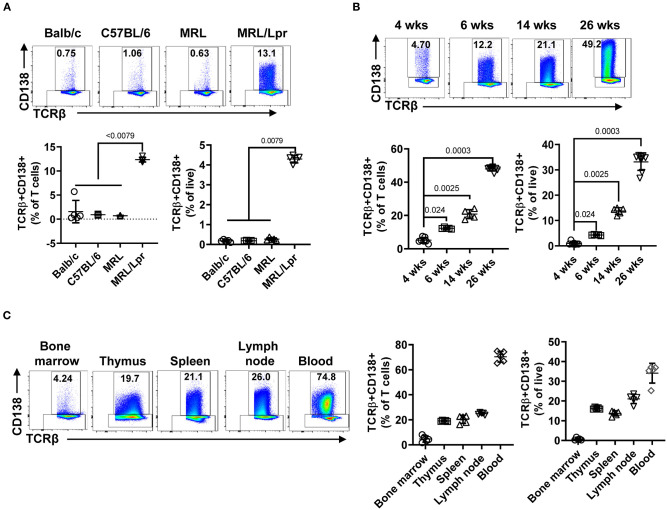
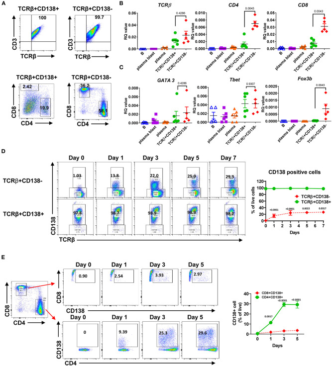
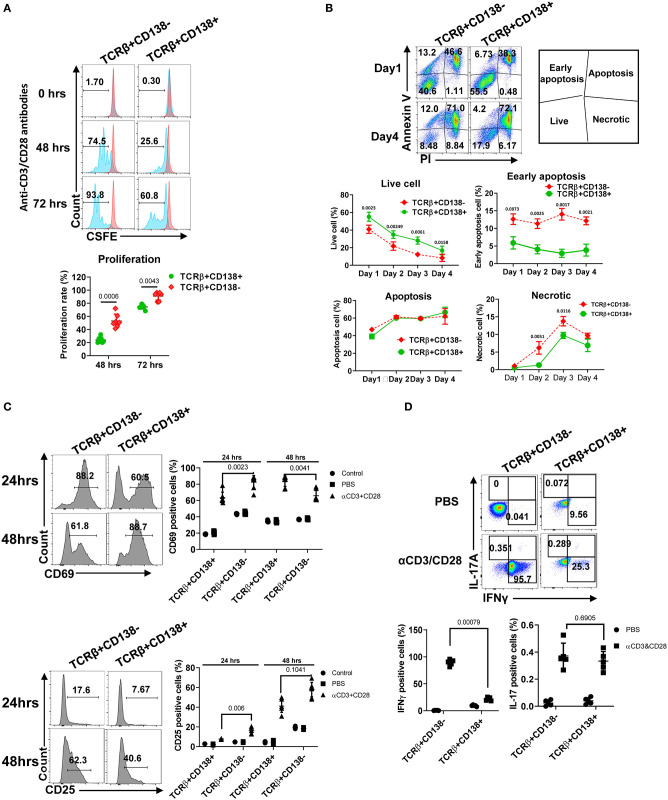
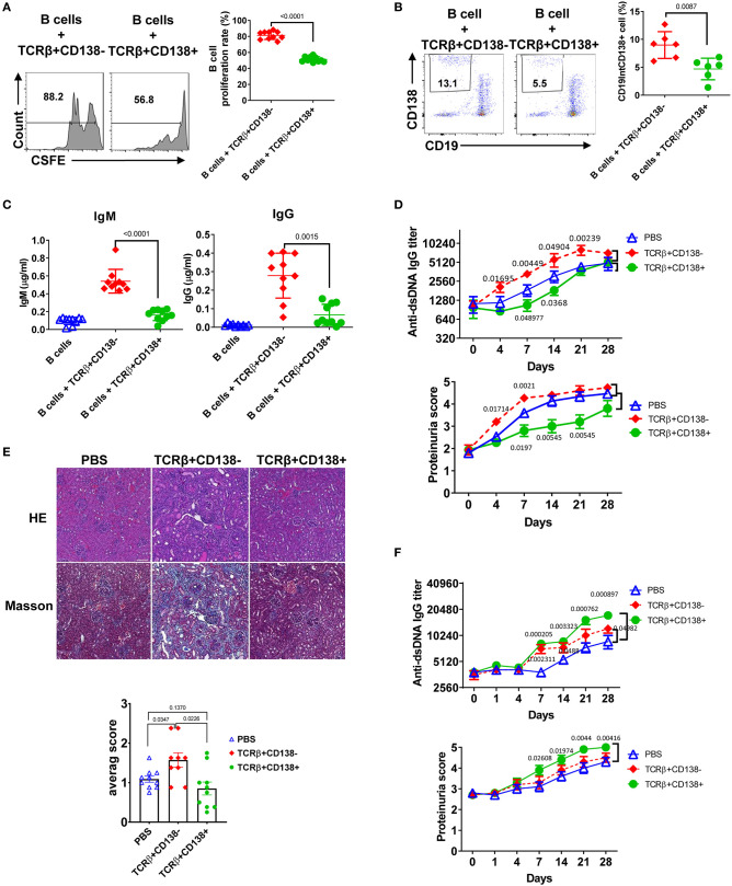
CD138 (syndecan 1), a member of the heparan-sulfate proteoglycan family, regulates diverse biological responses by interacting with chemokines, cytokines, growth factors, and adhesion molecules. Expression of CD138 has been detected on T cells from both healthy and sick mice mimicking systemic lupus erythematosus (SLE) disease. However, the characteristics and the role of CD138+ T cells in SLE pathogenesis remain largely unknown. We analyzed the lupus-prone MRL/Lpr mice and the control MRL/MpJ strain as well as the common laboratory strains Balb/c, and C57BL/6 for CD138-expression and found that only the MRL/Lpr strain harbored TCRβ+CD138+ cells in various organs. The frequency of TCRβ+CD138+ cells progressively expanded in MRL/Lpr mice with age and correlated with disease severity. Majority of the TCRβ+CD138+ cells were CD4 and CD8 double-negative and 20% were CD4. At least a portion of TCRβ+CD138+ cells originated from CD4+ cells because substantial number of CD4+TCRβ+CD138- cells expressed CD138 after cultivation. Compared to TCRβ+CD138- cells, TCRβ+CD138+ cells exhibited central memory (Tcm) phenotype with reduced ability to proliferate and produce the cytokines IFNγ and IL-17. When co-cultured with B cells, the ability of TCRβ+CD138+ cells to promote plasma cell formation and autoreactive antibody production was dependent on the presence of autoantigen, CD4 co-receptor expression and cell-to-cell contact. Surprisingly, adoptively transferred TCRβ+CD138+ T cells slowed down disease progression in young recipient MRL/Lpr mice but had the opposite effect when DNA was co-administered with TCRβ+CD138+ T cells or when TCRβ+CD138+ cells were transferred to older MRL/Lpr mice with established disease. Thus, CD138-expressing T cells with Tcm phenotype enhance disease progression in SLE by rapidly activating autoreactive B cells when self-antigens are exposed to the immune system.
CD138(黏附素 1)是硫酸乙酰肝素蛋白聚糖家族的成员,通过与趋化因子、细胞因子、生长因子和黏附分子相互作用,调节多种生物学反应。在模拟系统性红斑狼疮(SLE)疾病的健康和患病小鼠的 T 细胞中已经检测到 CD138 的表达。然而,CD138+T 细胞在 SLE 发病机制中的特征和作用在很大程度上仍然未知。我们分析了狼疮易感 MRL/Lpr 小鼠和对照 MRL/MpJ 品系以及常见的实验室品系 Balb/c 和 C57BL/6 中 CD138 的表达,发现只有 MRL/Lpr 品系在各种器官中存在 TCRβ+CD138+细胞。随着年龄的增长,MRL/Lpr 小鼠中 TCRβ+CD138+细胞的频率逐渐扩大,并与疾病严重程度相关。大多数 TCRβ+CD138+细胞为 CD4 和 CD8 双阴性,20%为 CD4+。至少一部分 TCRβ+CD138+细胞来源于 CD4+细胞,因为大量的 CD4+TCRβ+CD138-细胞在培养后表达 CD138。与 TCRβ+CD138-细胞相比,TCRβ+CD138+细胞表现出中央记忆(Tcm)表型,增殖能力和产生 IFNγ 和 IL-17 细胞因子的能力降低。当与 B 细胞共培养时,TCRβ+CD138+细胞促进浆细胞形成和自身反应性抗体产生的能力取决于自身抗原的存在、CD4 共受体表达和细胞间接触。令人惊讶的是,在年轻的受体 MRL/Lpr 小鼠中过继转移 TCRβ+CD138+T 细胞可减缓疾病进展,但当与 TCRβ+CD138+T 细胞共给予 DNA 或当 TCRβ+CD138+细胞转移到已建立疾病的老年 MRL/Lpr 小鼠时,效果相反。因此,具有 Tcm 表型的表达 CD138 的 T 细胞通过在免疫系统暴露于自身抗原时快速激活自身反应性 B 细胞,从而增强 SLE 中的疾病进展。