Liang Jingchen, Wang Duo, Li Hongxue, Zhao Shengfa, Chen Miao, Li Hang, Ding Zhanling, Liu Junjie, Liu Lianfeng
Department of Ultrasound, Affiliated Tumor Hospital of Guangxi Medical University, Nanning, Guangxi Zhuang Autonomous Region 530021, P.R. China.
Oncol Lett. 2020 Oct;20(4):75. doi: 10.3892/ol.2020.11936. Epub 2020 Jul 31.
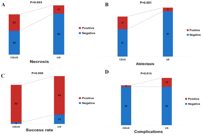
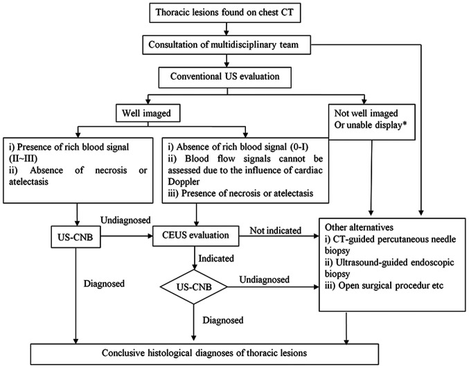
Two-dimensional ultrasound (US) and color doppler flow imaging are associated with certain limitations in the preprocedural evaluation and design of the puncture path for biopsies of thoracic lesions, such as a poorly defined boundary between the tumor and the atelectatic lesions in central lung cancer with atelectasis. Contrast-enhanced ultrasound (CEUS) can be valuable in the preoperative evaluation of the biopsy site and in increasing the accuracy of the biopsy. The present study investigated the value of clinical application of CEUS in US-guided core needle biopsy (US-CNB) in improving the diagnostic accuracy in thoracic lesions. A total of 120 patients with first-stage thoracic lesions from the Affiliated Tumor Hospital of Guangxi Medical University who underwent US-CNB were recruited and randomnly assigned to a conventional US group (n=66) and a CEUS group (n=54). All patients underwent preoperative evaluation and US-guided puncture of thoracic lesions. The intergroup differences in sonographic features, biopsy duration, biopsy success rate and complications were assessed. The CEUS group had a higher rate of detection of necrotic tissue (40.7% vs. 16.7%; χ=8.633; P=0.003) and change of initial puncture path (48.1%) compared with the US group. In central lung cancer with atelectasis, the ability to distinguish between tumor and atelectasis was higher in the CEUS group compared with the conventional US group (31.5 vs. 7.6%; χ=11.336; P=0.001). In addition, the CEUS group had a higher puncture success (96.3 vs. 80.3%; χ=6.946; P=0.008) and a lower complication rate (3.7% vs. 18.2%; χ=6.041; P=0.014) compared with the US group. CEUS can identify necrotic areas and occult tumors within atelectatic lung tissue and can be used for guiding puncture biopsy of thoracic lesions to improve the diagnostic accuracy with greater comparative clinical utility than conventional US. Pre-biopsy CEUS is especially useful for patients undergoing repeated US-CNB and those with hypovascular lesions, atelectasis or necrosis.
二维超声(US)和彩色多普勒血流成像在胸部病变活检的术前评估和穿刺路径设计方面存在一定局限性,例如中央型肺癌合并肺不张时肿瘤与肺不张病变之间边界不清。超声造影(CEUS)在活检部位的术前评估以及提高活检准确性方面可能具有重要价值。本研究探讨了CEUS在超声引导下经皮穿刺活检(US-CNB)中对提高胸部病变诊断准确性的临床应用价值。广西医科大学附属肿瘤医院共纳入120例接受US-CNB的I期胸部病变患者,并随机分为传统超声组(n = 66)和CEUS组(n = 54)。所有患者均接受术前评估及胸部病变的超声引导穿刺。评估两组在超声特征、活检持续时间、活检成功率及并发症方面的差异。与超声组相比,CEUS组坏死组织检出率更高(40.7% 对16.7%;χ = 8.633;P = 0.003),初始穿刺路径改变率更高(48.1%)。在中央型肺癌合并肺不张患者中,CEUS组区分肿瘤与肺不张的能力高于传统超声组(31.5对7.6%;χ = 11.336;P = 0.001)。此外,与超声组相比,CEUS组穿刺成功率更高(96.3%对80.3%;χ = 6.946;P = 0.008),并发症发生率更低(3.7%对18.2%;χ = 6.041;P = 0.014)。CEUS能够识别肺不张组织内的坏死区域和隐匿性肿瘤,可用于指导胸部病变的穿刺活检,与传统超声相比具有更大的临床比较实用性,能提高诊断准确性。活检前CEUS对接受重复US-CNB的患者以及血管少的病变、肺不张或坏死患者尤其有用。