Translational Research Center, Inha University, Incheon 22332, Korea.
Inha Research Institute for Medical Sciences, Inha University College of Medicine, Incheon 22332, Korea.
Int J Environ Res Public Health. 2020 Aug 31;17(17):6322. doi: 10.3390/ijerph17176322.
Most patients with thyroid cancer suffer from salivary gland (SG) dysfunctions after radioiodine (RI) therapy. We investigated the effects of keratinocyte growth factor (KGF)-1 on RI-induced SG dysfunction in an animal model.
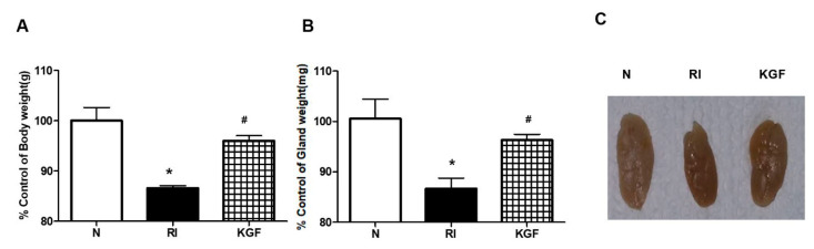
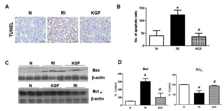
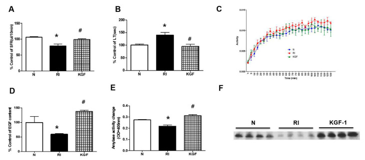
Six C57BL/6 mice were assigned to each of the following groups: treatment naïve control group, RI group, and RI+KGF-1 group. Body and SG weights, salivary flow rates, salivary lag times and changes in 99mTc pertechnetate uptake and excretion were measured, and histologic changes were noted. Amylase activities and epidermal growth factor (EGF) concentrations in saliva were also measured. In addition, TUNEL assays were performed and apoptosis-related protein expressions were assessed.
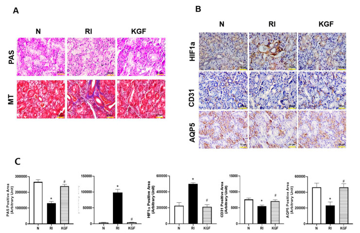
RI-induced reductions in salivary flow rates and increases in salivary lag times observed in the RI group were not observed in RI+KGF-1 group. Mice in RI group had higher HIF1a levels than controls, but HIF1a levels in RI+KGF-1 group were similar to those in control group. Furthermore, mice in RI+KGF-1 group had more mucin stained acini and decreased periductal fibrosis than mice in RI group, and tissue remodeling of many salivary epithelial cells (AQP5) and endothelial cells (CD31) were observed in RI+KGF-1 group. Amylase activity and expression in saliva were greater in RI+KGF-1 group than in RI group, and fewer apoptotic cells were observed in RI+KGF-1 group. Furthermore, BCLxl (anti-apoptotic) expression was higher, and Bax (pro-apoptotic) expression was lower in RI+KGF-1 group than in RI group.
Local delivery of KGF-1 might prevent RI-induced SG damage by reducing apoptosis.
大多数甲状腺癌患者在接受放射性碘(RI)治疗后会出现唾液腺(SG)功能障碍。我们在动物模型中研究了角质细胞生长因子(KGF)-1对 RI 诱导的 SG 功能障碍的影响。
将 6 只 C57BL/6 小鼠分别分配到以下各组:治疗初治对照组、RI 组和 RI+KGF-1 组。测量体重和 SG 重量、唾液流量、唾液滞后时间以及 99mTc 过锝酸盐摄取和排泄的变化,并观察组织学变化。还测量了唾液中的淀粉酶活性和表皮生长因子(EGF)浓度。此外,进行了 TUNEL 检测,并评估了凋亡相关蛋白的表达。
RI 组观察到的唾液流量减少和唾液滞后时间增加在 RI+KGF-1 组中未观察到。RI 组小鼠的 HIF1a 水平高于对照组,但 RI+KGF-1 组的 HIF1a 水平与对照组相似。此外,RI+KGF-1 组的小鼠有更多的粘蛋白染色的腺泡和减少的导管周围纤维化,比 RI 组的小鼠多,许多唾液上皮细胞(AQP5)和内皮细胞(CD31)的组织重塑在 RI+KGF-1 组中观察到。RI+KGF-1 组的唾液淀粉酶活性和表达高于 RI 组,且 RI+KGF-1 组的凋亡细胞较少。此外,RI+KGF-1 组的 BCLxl(抗凋亡)表达较高,Bax(促凋亡)表达较低。
局部递送 KGF-1 可能通过减少细胞凋亡来预防 RI 诱导的 SG 损伤。