Univ. of Michigan, United States.
J Biomed Opt. 2020 Sep;25(9). doi: 10.1117/1.JBO.25.9.095001.
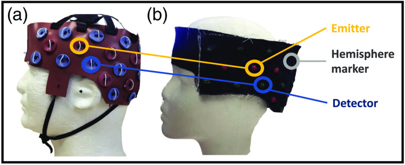
Functional near-infrared spectroscopy (fNIRS) is an emerging brain imaging technique due to its small size, low cost, minimum scanning sonic noise, and portability. Unfortunately, because this technique does not provide neuroanatomical information to accompany the functional data, its data interpretation remains a persistent challenge in fNIRS brain imaging applications. The two most popular approaches for fNIRS anatomical registration are magnetic resonance imaging (MRI) and three-dimensional (3-D) digitization. MRI scanning yields high-precision registration but reduces the cost-effectiveness and accessibility of fNIRS imaging. Alternatively, the low cost and portable 3-D digitizers are affected by magnetic properties of ambient metal objects, including participant clothing, testing equipment, medical implants, and so forth.
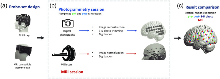
To overcome these obstacles and provide accessible and reliable neuroanatomical registration for fNIRS imaging, we developed and explored a photogrammetry optode registration (POR) method.
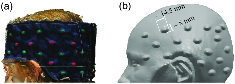
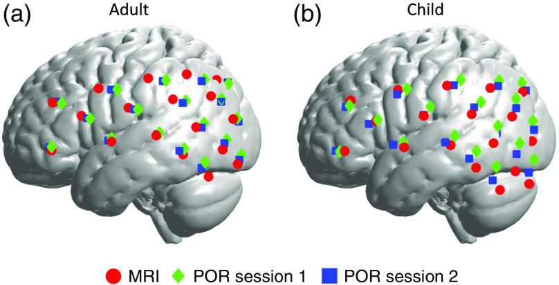
The POR method uses a consumer-grade camera to reconstruct a 3-D image of the fNIRS optode-set, including light emitters and detectors, on a participant's head. This reconstruction process uses a linear-time incremental structure from motion (LTI-SfM) algorithm, based on 100 to 150 digital photos. The POR method then aligns the reconstructed image with an anatomical template of the brain.
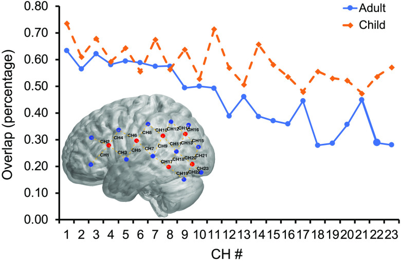
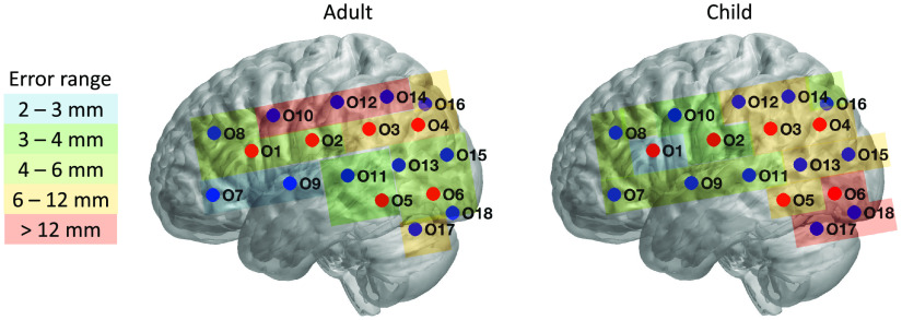
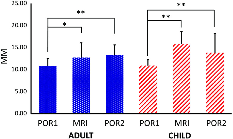
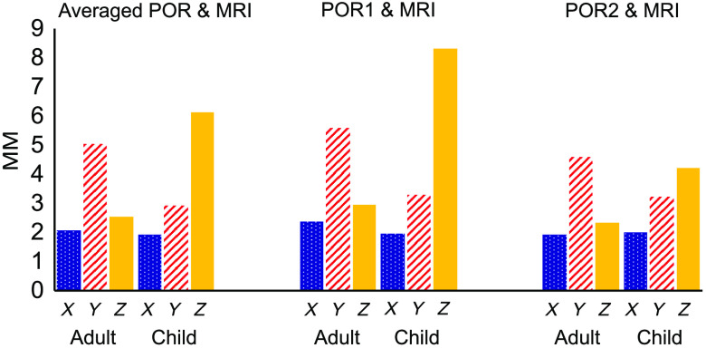
To validate this method, we tested 22 adult and 19 child participants using the POR method and MRI imaging. The results comparisons suggest on average 55% and 46% overlap across all data channel measurements registered by the two methods in adult and children, respectively. Importantly, this overlap reached 65% and 60% in only the frontal channels.
These results suggested that the mismatch in registration was partially due to higher variation in backward optode placement rather than the registration efficacy. Therefore, the photo-based registration method can offer an accessible and reliable approach to neuroanatomical registration of fNIRS as well as other surface-based neuroimaging and neuromodulation methods.
功能近红外光谱(fNIRS)是一种新兴的脑成像技术,因为它体积小、成本低、扫描噪音小且便携。不幸的是,由于该技术没有提供伴随功能数据的神经解剖信息,因此其数据解释仍然是 fNIRS 脑成像应用中的一个持续挑战。fNIRS 解剖配准最流行的两种方法是磁共振成像(MRI)和三维(3-D)数字化。MRI 扫描可实现高精度配准,但降低了 fNIRS 成像的成本效益和可及性。相反,低成本和便携式的 3-D 数字化仪受周围金属物体的磁性影响,包括参与者的衣物、测试设备、医疗植入物等。
为了克服这些障碍,并为 fNIRS 成像提供可及且可靠的神经解剖配准,我们开发并探索了一种摄影测量光极配准(POR)方法。
POR 方法使用消费级相机重建参与者头部上的 fNIRS 光极集的 3-D 图像,包括发射器和探测器。这个重建过程使用基于 100 到 150 张数码照片的线性时间增量结构从运动(LTI-SfM)算法。POR 方法然后将重建的图像与大脑的解剖模板对齐。
为了验证该方法,我们使用 POR 方法和 MRI 成像测试了 22 名成年参与者和 19 名儿童参与者。结果比较表明,在所有数据通道测量中,两种方法的平均重叠率分别为 55%和 46%,成人和儿童的重叠率分别为 65%和 60%,仅在额通道中。
这些结果表明,配准的不匹配部分是由于后向光极放置的变化较大,而不是配准效果。因此,基于照片的配准方法可以为 fNIRS 以及其他基于表面的神经影像学和神经调节方法提供一种可及且可靠的神经解剖配准方法。