Department of Radiology, Women's Hospital School of Medicine Zhejiang University, Hangzhou, 310006, Zhejiang province, China.
BMC Urol. 2020 Sep 4;20(1):142. doi: 10.1186/s12894-020-00708-0.
Primary vaginal calculus is rare and often misdiagnosed due to its low incidence. The formation of primary vaginal calculus is mainly due to the pooling and stasis of urine within the vagina, and associated with urogenital tract abnormalities.
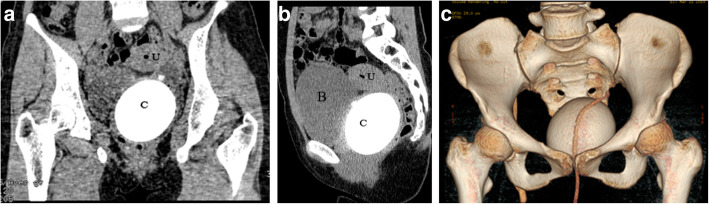
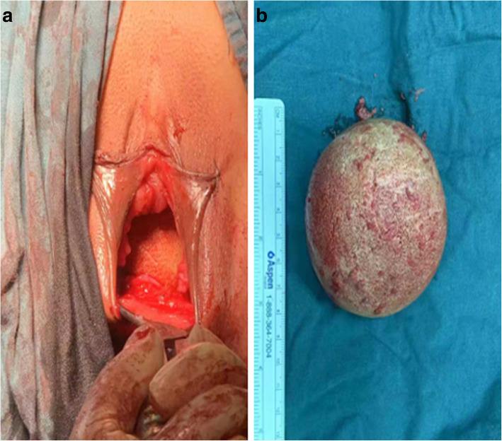
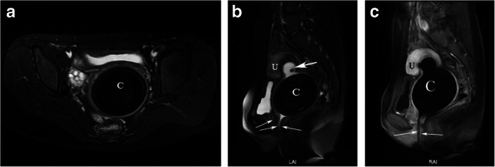
We present a case of a 23-year-old woman with urogenital sinus anomaly who presented with a vaginal calculus. The patient was not suspected of a calculus in the vagina until the patient suffered amenorrhea and dyspareunia. Pelvic computed tomography (CT) and magnetic resonance imaging (MRI) confirmed the patient had urogenital sinus anomaly with vaginal calculus. For the reason, the calculus was removed by surgery, and the reconstruction of vagina and urethra was performed. The postoperative recovery and follow-up were uneventful.
Although vaginal calculus and urogenital sinus anomaly are extremely rare in literature, the radiologist should be familiar with the imaging appearance of urogenital sinus anomaly, and be aware of the possibility of vaginal calculus.
原发性阴道结石罕见,由于其发病率低,常被误诊。原发性阴道结石的形成主要是由于尿液在阴道内积聚和停滞,并与泌尿生殖道异常有关。
我们报告了一例 23 岁女性,患有尿生殖窦畸形,表现为阴道结石。直到患者出现闭经和性交困难,才怀疑阴道内有结石。盆腔计算机断层扫描(CT)和磁共振成像(MRI)证实患者患有尿生殖窦畸形伴阴道结石。由于这个原因,通过手术切除了结石,并进行了阴道和尿道的重建。术后恢复和随访均顺利。
尽管阴道结石和尿生殖窦畸形在文献中极为罕见,但放射科医生应该熟悉尿生殖窦畸形的影像学表现,并意识到阴道结石的可能性。