• 文献检索
  • 文档翻译
  • 深度研究
  • 学术资讯
  • Suppr Zotero 插件Zotero 插件
  • 邀请有礼
  • 套餐&价格
  • 历史记录
应用&插件
Suppr Zotero 插件Zotero 插件浏览器插件Mac 客户端Windows 客户端微信小程序
定价
高级版会员购买积分包购买API积分包
服务
文献检索文档翻译深度研究API 文档MCP 服务
关于我们
关于 Suppr公司介绍联系我们用户协议隐私条款
关注我们

Suppr 超能文献

核心技术专利:CN118964589B侵权必究
粤ICP备2023148730 号-1Suppr @ 2026

文献检索

告别复杂PubMed语法,用中文像聊天一样搜索,搜遍4000万医学文献。AI智能推荐,让科研检索更轻松。

立即免费搜索

文件翻译

保留排版,准确专业,支持PDF/Word/PPT等文件格式,支持 12+语言互译。

免费翻译文档

深度研究

AI帮你快速写综述,25分钟生成高质量综述,智能提取关键信息,辅助科研写作。

立即免费体验

新冠肺炎(COVID-19)患者的基线胸部 X 光片:与临床和实验室数据的关联。

Baseline chest X-ray in coronavirus disease 19 (COVID-19) patients: association with clinical and laboratory data.

机构信息

Radiology Unit, Department of Surgical Sciences, University of Turin, Via Genova 3, 10126, Turin, Italy.

Radiology Unit, Department of Oncology, San Luigi Gonzaga Hospital, University of Turin, Orbassano, TO, Italy.

出版信息

Radiol Med. 2020 Dec;125(12):1271-1279. doi: 10.1007/s11547-020-01272-1. Epub 2020 Sep 7.

DOI:10.1007/s11547-020-01272-1
PMID:32894449
原文链接:https://pmc.ncbi.nlm.nih.gov/articles/PMC7475717/
Abstract

PURPOSE

To assess the reliability of CXR and to describe CXR findings and clinical and laboratory characteristics associated with positive and negative CXR.

METHODS

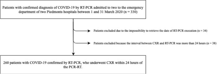
Retrospective two-center study on consecutive patients admitted to the emergency department of two north-western Italian hospitals in March 2020 with clinical suspicion of COVID-19 confirmed by RT-PCR and who underwent CXR within 24 h of the swab execution. 260 patients (61% male, 62.8 ± 15.8 year) were enrolled. CXRs were rated as positive (CXR+) or negative (CXR-), and features reported included presence and distribution of airspace opacities, pleural effusion and reduction in lung volumes. Clinical and laboratory data were collected. Statistical analysis was performed with nonparametric tests, binary logistic regression (BLR) and ROC curve analysis.

RESULTS

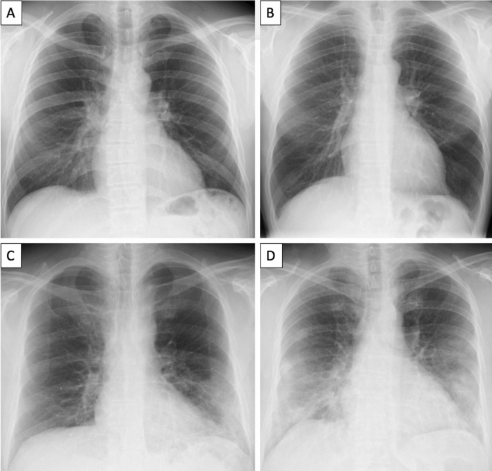
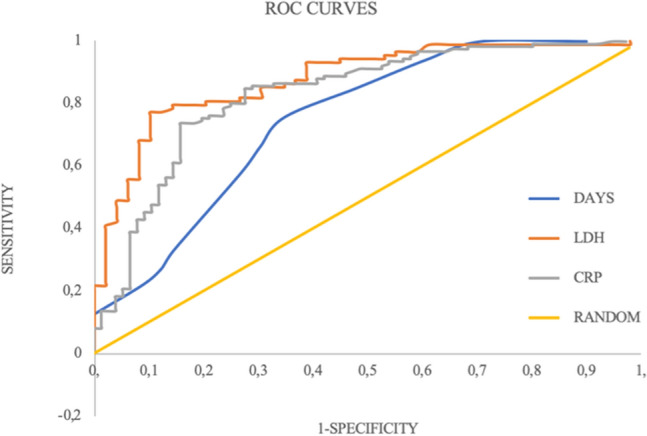
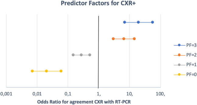
Sensitivity of CXR was 61.1% (95%CI 55-67%) with a typical presence of bilateral (62.3%) airspace opacification, more often with a lower zone (88.7%) and peripheral (43.4%) distribution. At univariate analysis, several factors were found to differ significantly between CXR+ and CXR-. The BLR confirmed as significant predictors only lactate dehydrogenase (LDH), C-reactive protein (CRP) and interval between the onset of symptoms and the execution of CXR. The ROC curve procedure determined that CRX+ was associated with LDH > 500 UI/L (AUC = 0.878), CRP > 30 mg/L (AUC = 0.830) and interval between the onset of symptoms and the execution of CXR > 4 days (AUC = 0.75). The presence of two out of three of the above-mentioned predictors resulted in CXR+ in 92.5% of cases, whereas their absence in 7.4%.

CONCLUSION

CXR has a low sensitivity. LDH, CRP and interval between the onset of symptoms and the execution of CXR are major predictors for a positive CXR.

摘要

目的

评估 CXR 的可靠性,并描述与 CXR 阳性和阴性相关的 CXR 结果和临床及实验室特征。

方法

这是一项回顾性的、在 2020 年 3 月于意大利西北部的两家医院急诊科进行的、连续纳入临床疑似 COVID-19 并经 RT-PCR 确诊且在拭子执行后 24 小时内接受 CXR 的患者的研究。共纳入 260 例患者(61%为男性,62.8±15.8 岁)。将 CXR 评为阳性(CXR+)或阴性(CXR-),并报告了空气腔隙混浊、胸腔积液和肺容积减少的存在和分布等特征。收集了临床和实验室数据。采用非参数检验、二项逻辑回归(BLR)和 ROC 曲线分析进行统计学分析。

结果

CXR 的敏感性为 61.1%(95%CI 55-67%),典型表现为双侧(62.3%)空气腔隙混浊,更常见于下区(88.7%)和外周(43.4%)分布。单因素分析发现,CXR+和 CXR-之间有几个因素存在显著差异。BLR 证实乳酸脱氢酶(LDH)、C 反应蛋白(CRP)和症状发作与 CXR 执行之间的间隔时间是唯一的显著预测因子。ROC 曲线程序确定 CXR+与 LDH>500 UI/L(AUC=0.878)、CRP>30 mg/L(AUC=0.830)和症状发作与 CXR 执行之间的间隔>4 天(AUC=0.75)相关。上述三个预测因子中有两个存在时,92.5%的情况下 CXR 为阳性,而不存在时为 7.4%。

结论

CXR 的敏感性较低。LDH、CRP 和症状发作与 CXR 执行之间的间隔时间是 CXR 阳性的主要预测因子。

https://cdn.ncbi.nlm.nih.gov/pmc/blobs/e0cc/7661418/bbb24dae5b19/11547_2020_1272_Fig4_HTML.jpg
https://cdn.ncbi.nlm.nih.gov/pmc/blobs/e0cc/7661418/ff42c5327b21/11547_2020_1272_Fig1_HTML.jpg
https://cdn.ncbi.nlm.nih.gov/pmc/blobs/e0cc/7661418/e09250210790/11547_2020_1272_Fig2_HTML.jpg
https://cdn.ncbi.nlm.nih.gov/pmc/blobs/e0cc/7661418/d198e4201a3b/11547_2020_1272_Fig3_HTML.jpg
https://cdn.ncbi.nlm.nih.gov/pmc/blobs/e0cc/7661418/bbb24dae5b19/11547_2020_1272_Fig4_HTML.jpg

相似文献

1
Baseline chest X-ray in coronavirus disease 19 (COVID-19) patients: association with clinical and laboratory data.新冠肺炎(COVID-19)患者的基线胸部 X 光片:与临床和实验室数据的关联。
Radiol Med. 2020 Dec;125(12):1271-1279. doi: 10.1007/s11547-020-01272-1. Epub 2020 Sep 7.
2
Chest X-ray in new Coronavirus Disease 2019 (COVID-19) infection: findings and correlation with clinical outcome.新型冠状病毒病 2019(COVID-19)感染的胸部 X 线表现:特征与临床转归的相关性。
Radiol Med. 2020 Aug;125(8):730-737. doi: 10.1007/s11547-020-01232-9. Epub 2020 Jun 9.
3
Rapid identification of SARS-CoV-2-infected patients at the emergency department using routine testing.在急诊科使用常规检测快速识别 SARS-CoV-2 感染患者。
Clin Chem Lab Med. 2020 Jun 29;58(9):1587-1593. doi: 10.1515/cclm-2020-0593. Print 2020 Aug 27.
4
Feasibility, Reproducibility, and Clinical Validity of a Quantitative Chest X-Ray Assessment for COVID-19.COVID-19 的定量胸部 X 射线评估的可行性、可重复性和临床有效性。
Am J Trop Med Hyg. 2020 Aug;103(2):822-827. doi: 10.4269/ajtmh.20-0535. Epub 2020 Jul 2.
5
Chest x-ray in the COVID-19 pandemic: Radiologists' real-world reader performance.COVID-19 大流行期间的胸部 X 光片:放射科医生的真实世界读者表现。
Eur J Radiol. 2020 Nov;132:109272. doi: 10.1016/j.ejrad.2020.109272. Epub 2020 Sep 10.
6
Thoracic imaging tests for the diagnosis of COVID-19.用于诊断新型冠状病毒肺炎的胸部影像学检查
Cochrane Database Syst Rev. 2020 Sep 30;9:CD013639. doi: 10.1002/14651858.CD013639.pub2.
7
Frequency and Distribution of Chest Radiographic Findings in Patients Positive for COVID-19.COVID-19 阳性患者的胸部 X 线表现的频率和分布。
Radiology. 2020 Aug;296(2):E72-E78. doi: 10.1148/radiol.2020201160. Epub 2020 Mar 27.
8
Correlation between Chest Computed Tomography and Lung Ultrasonography in Patients with Coronavirus Disease 2019 (COVID-19).新型冠状病毒肺炎(COVID-19)患者的胸部计算机断层扫描与肺部超声的相关性。
Ultrasound Med Biol. 2020 Nov;46(11):2918-2926. doi: 10.1016/j.ultrasmedbio.2020.07.003. Epub 2020 Jul 13.
9
Lactate dehydrogenase and C-reactive protein as predictors of respiratory failure in CoVID-19 patients.乳酸脱氢酶和 C 反应蛋白可预测 COVID-19 患者的呼吸衰竭。
Clin Chim Acta. 2020 Oct;509:135-138. doi: 10.1016/j.cca.2020.06.012. Epub 2020 Jun 9.
10
Chest CT in patients with a moderate or high pretest probability of COVID-19 and negative swab.胸部 CT 检查在中度或高度疑似 COVID-19 且咽拭子检测阴性的患者中的应用。
Radiol Med. 2020 Dec;125(12):1260-1270. doi: 10.1007/s11547-020-01269-w. Epub 2020 Aug 29.

引用本文的文献

1
Prognostic Value of the Brixia Radiological Score in COVID-19 Patients: A Retrospective Study from Romania.布丽夏放射学评分对新冠病毒肺炎患者的预后价值:罗马尼亚的一项回顾性研究
Trop Med Infect Dis. 2025 May 12;10(5):130. doi: 10.3390/tropicalmed10050130.
2
Clinical characteristics, outcomes, and costs of COVID-19 patients in Thai hospitels: a comparative analysis based on chest X-ray findings.泰国医院中新冠肺炎患者的临床特征、治疗结果及费用:基于胸部X光检查结果的比较分析
BMC Infect Dis. 2025 Jan 9;25(1):46. doi: 10.1186/s12879-025-10443-2.
3
Chest radiograph classification and severity of suspected COVID-19 by different radiologist groups and attending clinicians: multi-reader, multi-case study.

本文引用的文献

1
Lactate dehydrogenase levels predict coronavirus disease 2019 (COVID-19) severity and mortality: A pooled analysis.乳酸脱氢酶水平可预测 2019 冠状病毒病(COVID-19)的严重程度和死亡率:一项汇总分析。
Am J Emerg Med. 2020 Sep;38(9):1722-1726. doi: 10.1016/j.ajem.2020.05.073. Epub 2020 May 27.
2
Chest X-ray in new Coronavirus Disease 2019 (COVID-19) infection: findings and correlation with clinical outcome.新型冠状病毒病 2019(COVID-19)感染的胸部 X 线表现:特征与临床转归的相关性。
Radiol Med. 2020 Aug;125(8):730-737. doi: 10.1007/s11547-020-01232-9. Epub 2020 Jun 9.
3
Diagnostic impact of bedside chest X-ray features of 2019 novel coronavirus in the routine admission at the emergency department: case series from Lombardy region.
不同放射科医生组和临床医生对疑似 COVID-19 的胸部 X 线分类和严重程度:多读者、多病例研究。
Eur Radiol. 2023 Mar;33(3):2096-2104. doi: 10.1007/s00330-022-09172-w. Epub 2022 Oct 25.
4
Brixia Chest X-ray Severity Scoring System is in Relation with C-reactive Protein and D-dimer Values in Patients with COVID-19.布rixia胸部X光严重程度评分系统与新冠肺炎患者的C反应蛋白和D-二聚体值的关系
Mater Sociomed. 2022 Jun;34(2):95-99. doi: 10.5455/msm.2022.34.95-99.
5
Imaging Severity COVID-19 Assessment in Vaccinated and Unvaccinated Patients: Comparison of the Different Variants in a High Volume Italian Reference Center.接种疫苗和未接种疫苗患者的COVID-19影像严重程度评估:意大利一家大型参考中心不同变体的比较
J Pers Med. 2022 Jun 10;12(6):955. doi: 10.3390/jpm12060955.
6
Usefulness of Hospital Admission Chest X-ray Score for Predicting Mortality and ICU Admission in COVID-19 Patients.医院入院胸部X线评分对预测COVID-19患者死亡率和入住重症监护病房的价值
J Clin Med. 2022 Jun 20;11(12):3548. doi: 10.3390/jcm11123548.
7
Evaluation of Diagnostic Strategies for Identifying SARS-CoV-2 Infection in Clinical Practice: a Systematic Review and Compliance with the Standards for Reporting Diagnostic Accuracy Studies Guideline (STARD).评估临床实践中识别 SARS-CoV-2 感染的诊断策略:系统评价及对诊断准确性研究报告标准(STARD)的遵循情况。
Microbiol Spectr. 2022 Aug 31;10(4):e0030022. doi: 10.1128/spectrum.00300-22. Epub 2022 Jun 14.
8
Chest X-ray Severity Score as a Putative Predictor of Clinical Outcome in Hospitalized Patients: An Experience From a Vietnamese COVID-19 Field Hospital.胸部X光严重程度评分作为住院患者临床结局的一种假定预测指标:来自越南新冠肺炎野战医院的经验
Cureus. 2022 Mar 19;14(3):e23323. doi: 10.7759/cureus.23323. eCollection 2022 Mar.
9
Pulmonary Lymphangitis Poses a Major Challenge for Radiologists in an Oncological Setting during the COVID-19 Pandemic.在新冠疫情期间,肺部淋巴管炎给肿瘤学领域的放射科医生带来了重大挑战。
J Pers Med. 2022 Apr 12;12(4):624. doi: 10.3390/jpm12040624.
10
Systemic Emergencies in COVID-19 Patient: A Pictorial Review.新型冠状病毒肺炎患者的全身急危象:影像学综述。
Tomography. 2022 Apr 6;8(2):1041-1051. doi: 10.3390/tomography8020084.
2019 年新型冠状病毒在急诊科常规入院时床边胸部 X 线特征的诊断影响:来自伦巴第地区的病例系列。
Eur J Radiol. 2020 Aug;129:109092. doi: 10.1016/j.ejrad.2020.109092. Epub 2020 May 26.
4
Diagnostic Performance of Chest X-Ray for COVID-19 Pneumonia During the SARS-CoV-2 Pandemic in Lombardy, Italy.胸部 X 光在 SARS-CoV-2 大流行期间用于诊断意大利伦巴第 COVID-19 肺炎的性能。
J Thorac Imaging. 2020 Jul;35(4):W105-W106. doi: 10.1097/RTI.0000000000000533.
5
COVID-19 on Chest Radiographs: A Multireader Evaluation of an Artificial Intelligence System.COVID-19 胸片:人工智能系统的多读者评估。
Radiology. 2020 Sep;296(3):E166-E172. doi: 10.1148/radiol.2020201874. Epub 2020 May 8.
6
Clinical characteristics of coronavirus disease (COVID-19) early findings from a teaching hospital in Pavia, North Italy, 21 to 28 February 2020.2020 年 2 月 21 日至 28 日,意大利北部帕维亚教学医院对冠状病毒病(COVID-19)的早期发现的临床特征。
Euro Surveill. 2020 Apr;25(16). doi: 10.2807/1560-7917.ES.2020.25.16.2000460.
7
Portable chest X-ray in coronavirus disease-19 (COVID-19): A pictorial review.便携式胸部 X 光在冠状病毒病 19(COVID-19)中的应用:影像学综述。
Clin Imaging. 2020 Aug;64:35-42. doi: 10.1016/j.clinimag.2020.04.001. Epub 2020 Apr 8.
8
The Role of Chest Imaging in Patient Management during the COVID-19 Pandemic: A Multinational Consensus Statement from the Fleischner Society.《COVID-19 大流行期间患者管理中的胸部影像学作用:Fleischner 学会的一项多国共识声明》。
Radiology. 2020 Jul;296(1):172-180. doi: 10.1148/radiol.2020201365. Epub 2020 Apr 7.
9
Chest CT Features of COVID-19 in Rome, Italy.意大利罗马地区 COVID-19 的胸部 CT 特征。
Radiology. 2020 Aug;296(2):E79-E85. doi: 10.1148/radiol.2020201237. Epub 2020 Apr 3.
10
Frequency and Distribution of Chest Radiographic Findings in Patients Positive for COVID-19.COVID-19 阳性患者的胸部 X 线表现的频率和分布。
Radiology. 2020 Aug;296(2):E72-E78. doi: 10.1148/radiol.2020201160. Epub 2020 Mar 27.