• 文献检索
  • 文档翻译
  • 深度研究
  • 学术资讯
  • Suppr Zotero 插件Zotero 插件
  • 邀请有礼
  • 套餐&价格
  • 历史记录
应用&插件
Suppr Zotero 插件Zotero 插件浏览器插件Mac 客户端Windows 客户端微信小程序
定价
高级版会员购买积分包购买API积分包
服务
文献检索文档翻译深度研究API 文档MCP 服务
关于我们
关于 Suppr公司介绍联系我们用户协议隐私条款
关注我们

Suppr 超能文献

核心技术专利:CN118964589B侵权必究
粤ICP备2023148730 号-1Suppr @ 2026

文献检索

告别复杂PubMed语法,用中文像聊天一样搜索,搜遍4000万医学文献。AI智能推荐,让科研检索更轻松。

立即免费搜索

文件翻译

保留排版,准确专业,支持PDF/Word/PPT等文件格式,支持 12+语言互译。

免费翻译文档

深度研究

AI帮你快速写综述,25分钟生成高质量综述,智能提取关键信息,辅助科研写作。

立即免费体验

光学相干断层扫描血管造影黄斑灌注密度测量在周边视野前和周边视野青光眼的不对称性分析。

Asymmetry analysis of optical coherence tomography angiography macular perfusion density measurements in preperimetric and perimetric glaucoma.

机构信息

Department of Ophthalmology, Far Eastern Memorial Hospital, Ban-Chiao, New Taipei City, Taiwan, ROC.

Department of Medicine, National Taiwan University Hospital, Taipei, Taiwan, ROC.

出版信息

Sci Rep. 2020 Sep 8;10(1):14781. doi: 10.1038/s41598-020-71757-6.

DOI:10.1038/s41598-020-71757-6
PMID:32901074
原文链接:https://pmc.ncbi.nlm.nih.gov/articles/PMC7479593/
Abstract

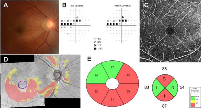
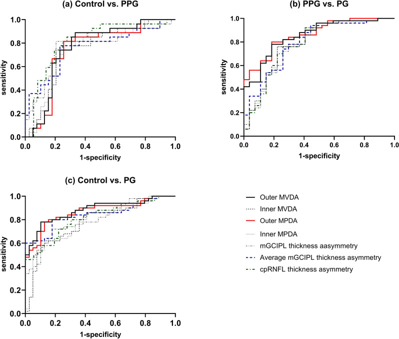
Macular retinal layer thickness asymmetry indices, particularly for the ganglion cell layer, are promising early indicators of glaucomatous damage. We evaluated macular perfusion density asymmetry (MPDA) among normal, preperimetric glaucoma (PPG), and perimetric glaucoma (PG) eyes, and we tested the performance of MPDA in differentiating between control and glaucoma eyes with or without visual field (VF) defects. In this study, 116 eyes (39 normal, 27 PPG, and 50 PG eyes) with optical coherence tomography angiography images of the macula were analysed. No significant difference was found in outer and inner MPDA between the control and PPG groups. However, outer MPDA was significantly higher in the PG group than in the PPG group (p = 0.009). Asymmetry of perfusion density and structural parameters was compared; no significant difference was found between controls and glaucoma patients. Outer MPDA had significantly higher discrimination ability between PPG and PG than did macular ganglion cell layer-inner plexiform layer thickness asymmetry (p = 0.039). In conclusion, the discriminant capability of MPDA for discriminating between glaucoma patients with and without VF defects is significantly higher than that of structural asymmetry. MPDA may be helpful in monitoring glaucoma progression in clinical practice.

摘要

黄斑视网膜层厚度不对称指数,特别是神经节细胞层,是青光眼损伤的有前途的早期指标。我们评估了正常、青光眼前期(PPG)和青光眼(PG)眼中的黄斑灌注密度不对称(MPDA),并测试了 MPDA 在区分有或无视野(VF)缺损的正常和青光眼眼中的表现。在这项研究中,分析了 116 只眼(39 只正常眼、27 只 PPG 眼和 50 只 PG 眼)的黄斑光学相干断层扫描血管造影图像。在对照组和 PPG 组之间,外和内 MPDA 没有显著差异。然而,PG 组的外 MPDA 明显高于 PPG 组(p=0.009)。比较了灌注密度和结构参数的不对称性;对照组和青光眼患者之间没有显著差异。外 MPDA 在区分 PPG 和 PG 方面的辨别能力明显高于黄斑神经节细胞层-内丛状层厚度不对称(p=0.039)。总之,MPDA 区分有和无 VF 缺损的青光眼患者的判别能力明显高于结构不对称。MPDA 可能有助于在临床实践中监测青光眼的进展。

https://cdn.ncbi.nlm.nih.gov/pmc/blobs/18f3/7479593/47054b74c07d/41598_2020_71757_Fig2_HTML.jpg
https://cdn.ncbi.nlm.nih.gov/pmc/blobs/18f3/7479593/2714e5f39864/41598_2020_71757_Fig1_HTML.jpg
https://cdn.ncbi.nlm.nih.gov/pmc/blobs/18f3/7479593/47054b74c07d/41598_2020_71757_Fig2_HTML.jpg
https://cdn.ncbi.nlm.nih.gov/pmc/blobs/18f3/7479593/2714e5f39864/41598_2020_71757_Fig1_HTML.jpg
https://cdn.ncbi.nlm.nih.gov/pmc/blobs/18f3/7479593/47054b74c07d/41598_2020_71757_Fig2_HTML.jpg

相似文献

1
Asymmetry analysis of optical coherence tomography angiography macular perfusion density measurements in preperimetric and perimetric glaucoma.光学相干断层扫描血管造影黄斑灌注密度测量在周边视野前和周边视野青光眼的不对称性分析。
Sci Rep. 2020 Sep 8;10(1):14781. doi: 10.1038/s41598-020-71757-6.
2
Structural evaluation of preperimetric and perimetric glaucoma.青光眼的视野前期和视野期的结构评估。
Indian J Ophthalmol. 2019 Nov;67(11):1843-1849. doi: 10.4103/ijo.IJO_1955_18.
3
Diagnostic ability of macular ganglion cell asymmetry in Preperimetric Glaucoma.黄斑神经节细胞不对称性在青光眼前期视野检查中的诊断能力
BMC Ophthalmol. 2019 Jan 8;19(1):12. doi: 10.1186/s12886-018-1019-4.
4
Rates and patterns of macular and circumpapillary retinal nerve fiber layer thinning in preperimetric and perimetric glaucomatous eyes.视野检查前和视野检查时青光眼患者黄斑及视乳头周围视网膜神经纤维层变薄的发生率和模式
J Glaucoma. 2015 Apr-May;24(4):278-85. doi: 10.1097/IJG.0000000000000046.
5
Ganglion cell-inner plexiform layer thickness of high definition optical coherence tomography in perimetric and preperimetric glaucoma.高分辨率光学相干断层扫描在视野缺损性青光眼和早期青光眼患者中的神经节细胞-内丛状层厚度研究
Invest Ophthalmol Vis Sci. 2014 Jul 11;55(8):4768-75. doi: 10.1167/iovs.14-14598.
6
Diagnostic Ability of Wide-field Retinal Nerve Fiber Layer Maps Using Swept-Source Optical Coherence Tomography for Detection of Preperimetric and Early Perimetric Glaucoma.使用扫频光学相干断层扫描技术的广角视网膜神经纤维层图谱对视野缺损前和早期视野缺损性青光眼的诊断能力
J Glaucoma. 2017 Jun;26(6):577-585. doi: 10.1097/IJG.0000000000000662.
7
Cluster analyses of grid-pattern display in macular parameters using optical coherence tomography for glaucoma diagnosis.利用光学相干断层扫描对青光眼诊断的黄斑参数进行网格图案显示的聚类分析。
Invest Ophthalmol Vis Sci. 2013 Sep 27;54(9):6401-8. doi: 10.1167/iovs.13-12805.
8
Glaucoma Detection Ability of Macular Ganglion Cell-Inner Plexiform Layer Thickness in Myopic Preperimetric Glaucoma.黄斑神经节细胞-内丛状层厚度在近视性视野缺损前期青光眼患者中的青光眼检测能力
Invest Ophthalmol Vis Sci. 2015 Dec;56(13):8306-13. doi: 10.1167/iovs.15-18141.
9
Macular vessel density versus ganglion cell complex thickness for detection of early primary open-angle glaucoma.黄斑血管密度与神经节细胞复合体厚度在早期原发性开角型青光眼检测中的比较。
BMC Ophthalmol. 2020 Jan 8;20(1):17. doi: 10.1186/s12886-020-1304-x.
10
Macular Ganglion Cell Layer Assessment to Detect Glaucomatous Central Visual Field Progression.用于检测青光眼性中心视野进展的黄斑神经节细胞层评估
Korean J Ophthalmol. 2016 Dec;30(6):451-458. doi: 10.3341/kjo.2016.30.6.451. Epub 2016 Dec 6.

引用本文的文献

1
Macular microvasculature asymmetry in high myopia patients with early open-angle glaucoma.早期开角型青光眼高度近视患者的黄斑部微血管不对称性
Sci Rep. 2025 Jul 2;15(1):22928. doi: 10.1038/s41598-025-06774-4.
2
Innovative Macula Capillaries Plexuses Visualization with OCTA B-Scan Graph Representation: Transforming OCTA B-Scan into OCTA Graph Representation.利用光学相干断层扫描血管造影(OCTA)B 扫描图表示法实现创新的黄斑毛细血管丛可视化:将 OCTA B 扫描转换为 OCTA 图表示法。
J Multidiscip Healthc. 2023 Nov 15;16:3477-3491. doi: 10.2147/JMDH.S433405. eCollection 2023.
3
The Diagnostic Value of Pulsar Perimetry, Optical Coherence Tomography, and Optical Coherence Tomography Angiography in Pre-Perimetric and Perimetric Glaucoma.
脉冲星视野检查、光学相干断层扫描及光学相干断层扫描血管造影在周边视野前及周边视野青光眼诊断中的价值
J Clin Med. 2021 Dec 13;10(24):5825. doi: 10.3390/jcm10245825.
4
Vessel Density in the Macular and Peripapillary Areas in Preperimetric Glaucoma to Various Stages of Primary Open-Angle Glaucoma in Taiwan.台湾青光眼前期黄斑区和视乳头周围区域血管密度与原发性开角型青光眼各阶段的关系
J Clin Med. 2021 Nov 23;10(23):5490. doi: 10.3390/jcm10235490.