Cleven A H G, Hanff D F, Hartgrink H, Dijkstra P D S
Department of Orthopaedic and Traumatology, Adam Malik General Hospital / Faculty of Medicine Universitas Sumatera Utara, Medan, Indonesia.
Department of Pathology, Leiden University Medical Center, Leiden, the Netherlands.
Ann Med Surg (Lond). 2020 Aug 2;57:274-280. doi: 10.1016/j.amsu.2020.07.002. eCollection 2020 Sep.
Myelolipomas are very rare benign tumours consisting of hematopoietic cells and mature adipose tissues. They are most commonly found in the adrenal glands. However, there have been several reported cases of extra-adrenal myelolipomas, most commonly in the presacral region. Nearly all presacral lesions are small and asymptomatic; thus, most are discovered incidentally on imaging studies.
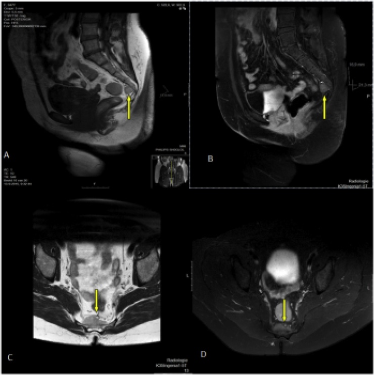
We report two cases of presacral myelolipomas. The first is a 48-year-old female presenting with atypical back pain, found to have a mass in her presacral region with a size of 3,3 cm. The second case is a 59-year-old female, who presented for evaluation of a hip fracture, found to have a 4,7 cm presacral lesion. Both presacral myelolipomas were discovered incidentally and were confirmed by percutaneous guided fine-needle aspiration biopsy. Both were treated conservatively.
Accepted indications for the surgical excision of myelolipomas are symptomatic tumour, size >4 cm, metabolically active tumour, and a suspicion of malignancy on an imaging study. However, previous reports have documented that nearly half of the conservatively managed myelolipomas with a mean initial size of 5,1 cm, has increased in size or became symptomatic over a 3-years period.
We conclude that symptomatic presacral myelolipomas or lesions larger than 4 cm should be en-bloc resected, and we present an intuitive decision-making algorithm.
髓脂肪瘤是由造血细胞和成熟脂肪组织构成的极为罕见的良性肿瘤。它们最常出现在肾上腺。然而,已有数例肾上腺外髓脂肪瘤的报道,最常见于骶前区域。几乎所有骶前病变都较小且无症状;因此,大多数是在影像学检查时偶然发现的。
我们报告两例骶前髓脂肪瘤。第一例是一名48岁女性,表现为非典型背痛,发现骶前区域有一个大小为3.3厘米的肿块。第二例是一名59岁女性,因髋部骨折前来评估,发现有一个4.7厘米的骶前病变。这两例骶前髓脂肪瘤均为偶然发现,并经皮引导细针穿刺活检确诊。两例均采用保守治疗。
髓脂肪瘤手术切除的公认指征是有症状的肿瘤、大小>4厘米、代谢活跃的肿瘤以及影像学检查怀疑为恶性。然而,既往报道显示,平均初始大小为5.1厘米的保守治疗的髓脂肪瘤中,近一半在3年期间大小增加或出现症状。
我们得出结论,有症状的骶前髓脂肪瘤或大于4厘米的病变应整块切除,并且我们提出了一种直观的决策算法。