Nuclear Medicine Unit, Humanitas Gavazzeni, Bergamo, Italy.
Radiology Unit, Humanitas Gavazzeni, Bergamo, Italy.
Eur J Nucl Med Mol Imaging. 2021 Mar;48(3):777-785. doi: 10.1007/s00259-020-05027-y. Epub 2020 Sep 9.
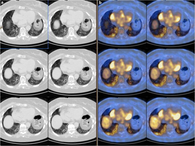
The study aimed to compare the incidence of interstitial pneumonia on [F]-FDG PET/CT scans between two 6-month periods: (a) the COVID-19 pandemic peak and (b) control period. Secondly, we compared the incidence of interstitial pneumonia on [F]-FDG PET/CT and epidemiological data from the regional registry of COVID-19 cases. Additionally, imaging findings and the intensity of [F]-FDG PET/CT uptake in terms of maximum standardized uptake value (SUVmax) were compared.
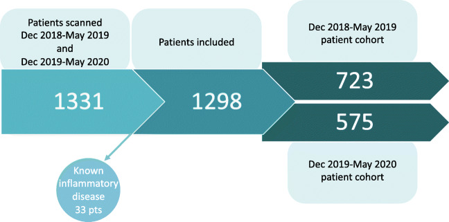
We retrospectively analyzed [F]-FDG PET/CT scans performed in cancer patients referred to nuclear medicine of Humanitas Gavazzeni in Bergamo from December 2019 to May 2020 and from December 2018 to May 2019. The per month incidence of interstitial pneumonia at imaging and the epidemiological data were assessed. To evaluate the differences between the two symmetric groups (period of COVID-19 pandemic and control), the stratified Cochran-Mantel-Haenszel test was used. Chi-square test or Fisher's exact test and t test or Wilcoxon test were performed to compare the distributions of categorical and continuous variables, respectively.
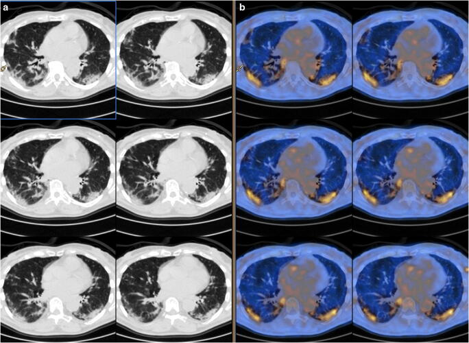
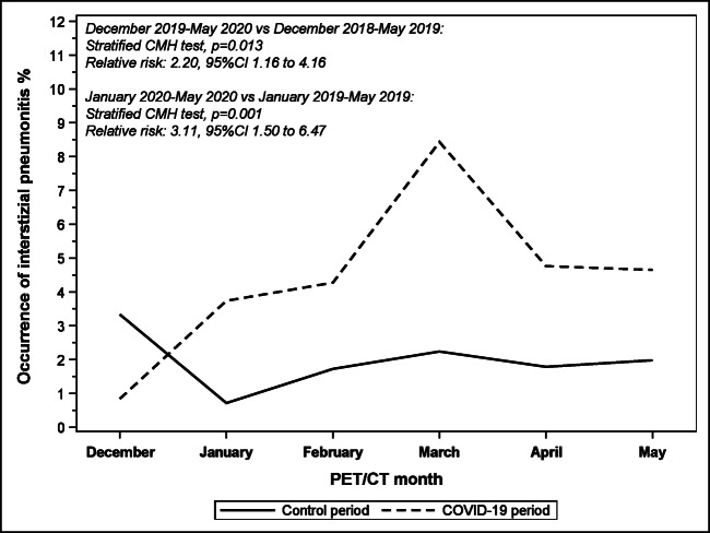
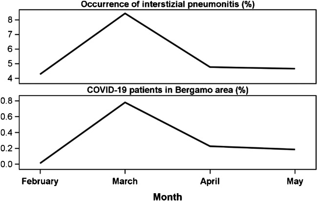
Overall, 1298 patients were included in the study. The two cohorts-COVID-19 pandemic (n = 575) and control (n = 723)-did not statistically differ in terms of age, disease, or scan indication (p > 0.05). Signs of interstitial pneumonia were observed in 24 (4.2%) and 14 patients (1.9%) in the COVID-19 period and the control period, respectively, with a statistically significant difference (p = 0.013). The level of statistical significance improved further when the period from January to May was considered, with a peak in March (7/83 patients, 8.4% vs 3/134 patients, 2.2%, p = 0.001). The curve of interstitial pneumonia diagnosis overlapped with the COVID-19 incidence in the area of Lombardy (Spearman correlation index was equal to 1). Imaging data did not differ among the two cohorts.
Significant increase of interstitial lung alterations at [F]-FDG PET/CT has been demonstrated during the COVID-19 pandemic. Additionally, the incidence curve of imaging abnormalities resulted in resembling the epidemiological data of the general population. These data support the rationale to adopt [F]-FDG PET/CT as sentinel modality to identify suspicious COVID-19 cases to be referred for additional confirmatory testing. Nuclear medicine physicians and staff should continue active surveillance of interstitial pneumonia findings, especially when new infection peak is expected.
本研究旨在比较 COVID-19 大流行高峰期和对照期两个 6 个月期间[F]-FDG PET/CT 扫描中间质性肺炎的发生率。其次,我们比较了[F]-FDG PET/CT 扫描与 COVID-19 区域登记处的流行病学数据之间的间质性肺炎发生率。此外,还比较了影像学表现和最大标准化摄取值(SUVmax)方面[F]-FDG PET/CT 摄取的强度。
我们回顾性分析了 2019 年 12 月至 2020 年 5 月和 2018 年 12 月至 2019 年 5 月期间在 Bergamo 的 Humanitas Gavazzeni 核医学科就诊的癌症患者的[F]-FDG PET/CT 扫描。评估了影像学检查中每月间质性肺炎的发生率和流行病学数据。为了评估两个对称组(COVID-19 大流行期和对照组)之间的差异,使用分层 Cochran-Mantel-Haenszel 检验。卡方检验或 Fisher 确切检验和 t 检验或 Wilcoxon 检验分别用于比较分类变量和连续变量的分布。
总体而言,共有 1298 例患者纳入研究。COVID-19 大流行期间(n=575)和对照组(n=723)两组在年龄、疾病或扫描指征方面无统计学差异(p>0.05)。COVID-19 期间观察到 24 例(4.2%)和对照组 14 例(1.9%)患者存在间质性肺炎征象,差异有统计学意义(p=0.013)。当考虑从 1 月至 5 月的时间段时,统计学意义的水平进一步提高,3 月达到高峰(7/83 例,8.4%比 3/134 例,2.2%,p=0.001)。间质性肺炎诊断曲线与伦巴第地区 COVID-19 发病率重叠(Spearman 相关指数等于 1)。两组的影像学数据无差异。
在 COVID-19 大流行期间,[F]-FDG PET/CT 上的间质性肺改变明显增加。此外,影像学异常的发生率曲线与一般人群的流行病学数据相似。这些数据支持将[F]-FDG PET/CT 作为识别可疑 COVID-19 病例的哨点模态的合理性,这些病例需要进一步进行确认性检测。核医学医师和工作人员应继续积极监测间质性肺炎的发现,特别是当预计出现新的感染高峰时。