• 文献检索
  • 文档翻译
  • 深度研究
  • 学术资讯
  • Suppr Zotero 插件Zotero 插件
  • 邀请有礼
  • 套餐&价格
  • 历史记录
应用&插件
Suppr Zotero 插件Zotero 插件浏览器插件Mac 客户端Windows 客户端微信小程序
定价
高级版会员购买积分包购买API积分包
服务
文献检索文档翻译深度研究API 文档MCP 服务
关于我们
关于 Suppr公司介绍联系我们用户协议隐私条款
关注我们

Suppr 超能文献

核心技术专利:CN118964589B侵权必究
粤ICP备2023148730 号-1Suppr @ 2026

文献检索

告别复杂PubMed语法,用中文像聊天一样搜索,搜遍4000万医学文献。AI智能推荐,让科研检索更轻松。

立即免费搜索

文件翻译

保留排版,准确专业,支持PDF/Word/PPT等文件格式,支持 12+语言互译。

免费翻译文档

深度研究

AI帮你快速写综述,25分钟生成高质量综述,智能提取关键信息,辅助科研写作。

立即免费体验

2021年初新型冠状病毒肺炎影像学表现的综合综述——现状

A comprehensive review of imaging findings in COVID-19 - status in early 2021.

作者信息

Afshar-Oromieh Ali, Prosch Helmut, Schaefer-Prokop Cornelia, Bohn Karl Peter, Alberts Ian, Mingels Clemens, Thurnher Majda, Cumming Paul, Shi Kuangyu, Peters Alan, Geleff Silvana, Lan Xiaoli, Wang Feng, Huber Adrian, Gräni Christoph, Heverhagen Johannes T, Rominger Axel, Fontanellaz Matthias, Schöder Heiko, Christe Andreas, Mougiakakou Stavroula, Ebner Lukas

机构信息

Department of Nuclear Medicine, Inselspital, Bern University Hospital, University of Bern, Freiburgstr. 18, CH-3010, Bern, Switzerland.

Department of Biomedical Imaging and Image-guided Therapy, Medical University Vienna, Vienna, Austria.

出版信息

Eur J Nucl Med Mol Imaging. 2021 Jul;48(8):2500-2524. doi: 10.1007/s00259-021-05375-3. Epub 2021 May 1.

DOI:10.1007/s00259-021-05375-3
PMID:33932183
原文链接:https://pmc.ncbi.nlm.nih.gov/articles/PMC8087891/
Abstract

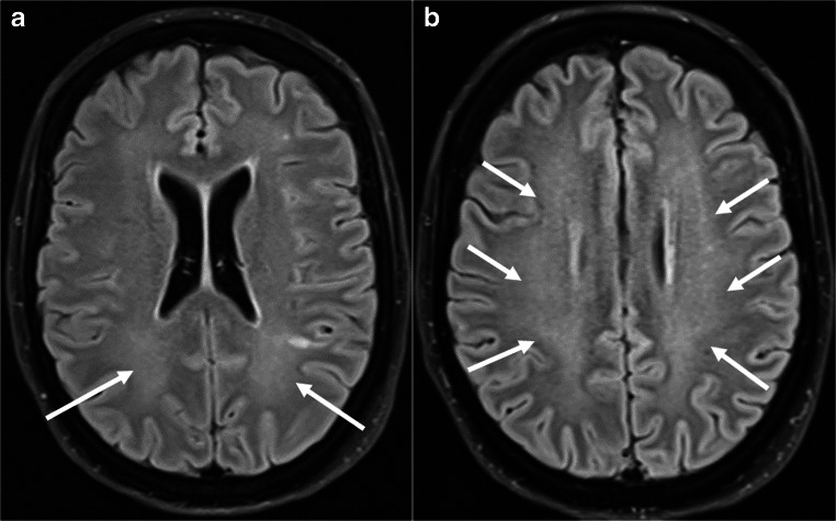
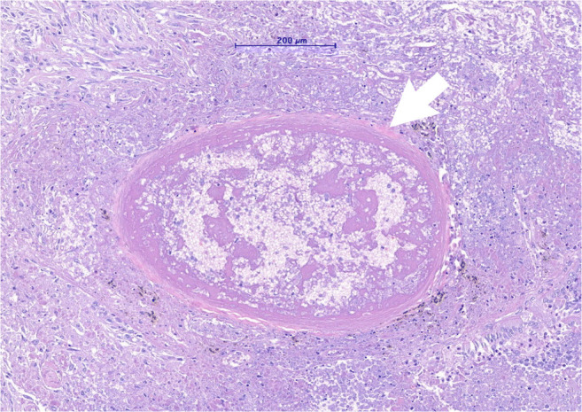
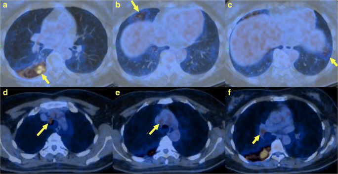
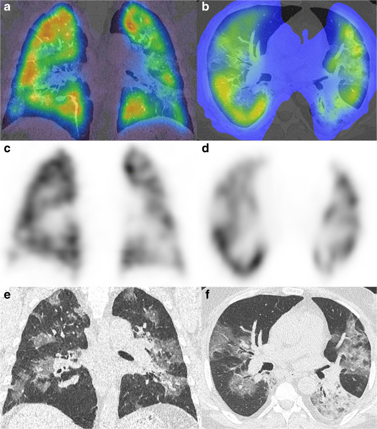
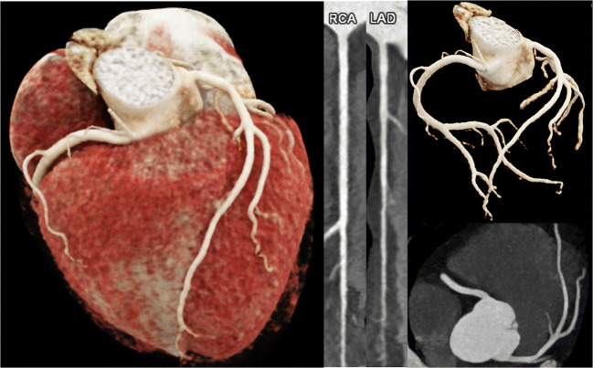
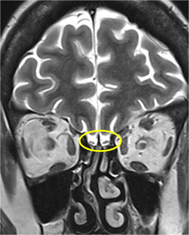
Medical imaging methods are assuming a greater role in the workup of patients with COVID-19, mainly in relation to the primary manifestation of pulmonary disease and the tissue distribution of the angiotensin-converting-enzyme 2 (ACE 2) receptor. However, the field is so new that no consensus view has emerged guiding clinical decisions to employ imaging procedures such as radiography, computer tomography (CT), positron emission tomography (PET), and magnetic resonance imaging, and in what measure the risk of exposure of staff to possible infection could be justified by the knowledge gained. The insensitivity of current RT-PCR methods for positive diagnosis is part of the rationale for resorting to imaging procedures. While CT is more sensitive than genetic testing in hospitalized patients, positive findings of ground glass opacities depend on the disease stage. There is sparse reporting on PET/CT with [F]-FDG in COVID-19, but available results are congruent with the earlier literature on viral pneumonias. There is a high incidence of cerebral findings in COVID-19, and likewise evidence of gastrointestinal involvement. Artificial intelligence, notably machine learning is emerging as an effective method for diagnostic image analysis, with performance in the discriminative diagnosis of diagnosis of COVID-19 pneumonia comparable to that of human practitioners.

摘要

医学成像方法在新型冠状病毒肺炎(COVID-19)患者的检查中发挥着越来越重要的作用,主要涉及肺部疾病的主要表现以及血管紧张素转换酶2(ACE 2)受体的组织分布。然而,该领域非常新,尚未形成共识观点来指导诸如放射照相、计算机断层扫描(CT)、正电子发射断层扫描(PET)和磁共振成像等成像程序的临床决策,以及通过所获得的知识来证明工作人员接触可能感染的风险在何种程度上是合理的。当前逆转录聚合酶链反应(RT-PCR)方法对阳性诊断的不敏感性是采用成像程序的部分理由。虽然CT在住院患者中比基因检测更敏感,但磨玻璃影的阳性结果取决于疾病阶段。关于COVID-19中[F]-FDG PET/CT的报道很少,但现有结果与早期关于病毒性肺炎的文献一致。COVID-19中脑部发现的发生率很高,同样也有胃肠道受累的证据。人工智能,尤其是机器学习正在成为诊断图像分析的有效方法,在COVID-19肺炎的鉴别诊断中的表现与人类从业者相当。

https://cdn.ncbi.nlm.nih.gov/pmc/blobs/09e1/8241739/7141cbab2aa2/259_2021_5375_Fig10_HTML.jpg
https://cdn.ncbi.nlm.nih.gov/pmc/blobs/09e1/8241739/2cd6d48699b6/259_2021_5375_Fig1_HTML.jpg
https://cdn.ncbi.nlm.nih.gov/pmc/blobs/09e1/8241739/9a497089d10c/259_2021_5375_Fig2_HTML.jpg
https://cdn.ncbi.nlm.nih.gov/pmc/blobs/09e1/8241739/a5c46b5df6b3/259_2021_5375_Fig3_HTML.jpg
https://cdn.ncbi.nlm.nih.gov/pmc/blobs/09e1/8241739/6af201a3a78f/259_2021_5375_Fig4_HTML.jpg
https://cdn.ncbi.nlm.nih.gov/pmc/blobs/09e1/8241739/23ffd8d9b859/259_2021_5375_Fig5_HTML.jpg
https://cdn.ncbi.nlm.nih.gov/pmc/blobs/09e1/8241739/8b610a22ae57/259_2021_5375_Fig6_HTML.jpg
https://cdn.ncbi.nlm.nih.gov/pmc/blobs/09e1/8241739/f2bd4d10018e/259_2021_5375_Fig7_HTML.jpg
https://cdn.ncbi.nlm.nih.gov/pmc/blobs/09e1/8241739/078a25497f7c/259_2021_5375_Fig8_HTML.jpg
https://cdn.ncbi.nlm.nih.gov/pmc/blobs/09e1/8241739/84ddf60bb551/259_2021_5375_Fig9_HTML.jpg
https://cdn.ncbi.nlm.nih.gov/pmc/blobs/09e1/8241739/7141cbab2aa2/259_2021_5375_Fig10_HTML.jpg

相似文献

1
A comprehensive review of imaging findings in COVID-19 - status in early 2021.2021年初新型冠状病毒肺炎影像学表现的综合综述——现状
Eur J Nucl Med Mol Imaging. 2021 Jul;48(8):2500-2524. doi: 10.1007/s00259-021-05375-3. Epub 2021 May 1.
2
Increased incidence of interstitial pneumonia detected on [F]-FDG-PET/CT in asymptomatic cancer patients during COVID-19 pandemic in Lombardy: a casualty or COVID-19 infection?在 COVID-19 大流行期间,伦巴第无症状癌症患者的 [F]-FDG-PET/CT 检测到间质性肺炎发病率增加:是 COVID-19 感染的偶然事件还是 COVID-19 感染?
Eur J Nucl Med Mol Imaging. 2021 Mar;48(3):777-785. doi: 10.1007/s00259-020-05027-y. Epub 2020 Sep 9.
3
F-FDG PET/CT findings of COVID-19: a series of four highly suspected cases.COVID-19 的 F-FDG PET/CT 表现:四例高度疑似病例系列报道。
Eur J Nucl Med Mol Imaging. 2020 May;47(5):1281-1286. doi: 10.1007/s00259-020-04734-w. Epub 2020 Feb 22.
4
[F]-FDG PET/CT in oncologic patients with unsuspected asymptomatic infection with SARS-CoV-2.[F]-FDG PET/CT 在疑似无症状感染 SARS-CoV-2 的肿瘤患者中的应用。
Eur J Nucl Med Mol Imaging. 2021 Mar;48(3):786-793. doi: 10.1007/s00259-020-04979-5. Epub 2020 Sep 16.
5
FDG-PET/CT findings highly suspicious for COVID-19 in an Italian case series of asymptomatic patients.在意大利一系列无症状患者中,FDG-PET/CT 检查结果高度疑似 COVID-19。
Eur J Nucl Med Mol Imaging. 2020 Jul;47(7):1649-1656. doi: 10.1007/s00259-020-04819-6. Epub 2020 Apr 27.
6
Unknown SARS-CoV-2 pneumonia detected by PET/CT in patients with cancer.PET/CT检测出癌症患者中的不明新型冠状病毒肺炎。
Tumori. 2020 Aug;106(4):325-332. doi: 10.1177/0300891620935983. Epub 2020 Jun 22.
7
Possible Alterations of Imaging Patterns in Computed Tomography for Delta-VOC of SARS-CoV-2.新冠病毒德尔塔变异株 CT 影像学特征变化
Rofo. 2022 Nov;194(11):1229-1241. doi: 10.1055/a-1826-0436. Epub 2022 Jul 18.
8
Similarities and Differences of Early Pulmonary CT Features of Pneumonia Caused by SARS-CoV-2, SARS-CoV and MERS-CoV: Comparison Based on a Systemic Review.新型冠状病毒、严重急性呼吸综合征冠状病毒和中东呼吸综合征冠状病毒所致肺炎早期肺部CT特征的异同:基于系统评价的比较
Chin Med Sci J. 2020 Sep 30;35(3):254-261. doi: 10.24920/003727.
9
Imaging and clinical features of patients with 2019 novel coronavirus SARS-CoV-2.新型冠状病毒 2019 年 SARS-CoV-2 患者的影像学和临床特征。
Eur J Nucl Med Mol Imaging. 2020 May;47(5):1275-1280. doi: 10.1007/s00259-020-04735-9. Epub 2020 Feb 28.
10
Incidental Findings Suggestive of COVID-19 Pneumonia in Oncologic Patients Undergoing F-FDG PET/CT Studies: Association Between Metabolic and Structural Lung Changes.在接受 F-FDG PET/CT 研究的肿瘤患者中提示 COVID-19 肺炎的偶然发现:代谢和结构性肺改变之间的关联。
J Nucl Med. 2022 Feb;63(2):274-279. doi: 10.2967/jnumed.121.261915. Epub 2021 Jun 4.

引用本文的文献

1
Association of High-Risk Obstructive Sleep Apnea with Artificial Intelligence-Guided, CT-Based Severity Scores in Patients with COVID-19 Pneumonia.高危阻塞性睡眠呼吸暂停与基于CT的人工智能指导的COVID-19肺炎患者严重程度评分的关联
J Clin Med. 2024 Oct 26;13(21):6415. doi: 10.3390/jcm13216415.
2
Molecular Imaging of ACE2 Expression in Infectious Disease and Cancer.在传染病和癌症中 ACE2 表达的分子成像。
Viruses. 2023 Sep 23;15(10):1982. doi: 10.3390/v15101982.
3
Diagnosis, treatment and prevention of severe acute respiratory syndrome coronavirus 2 infection in children: experts' consensus statement updated for the Omicron variant.

本文引用的文献

1
Automated Assessment and Tracking of COVID-19 Pulmonary Disease Severity on Chest Radiographs using Convolutional Siamese Neural Networks.使用卷积暹罗神经网络对胸部X光片上的COVID-19肺部疾病严重程度进行自动评估和跟踪。
Radiol Artif Intell. 2020 Jul 22;2(4):e200079. doi: 10.1148/ryai.2020200079. eCollection 2020 Jul.
2
Pulmonary Thromboembolism in COVID-19: Venous Thromboembolism or Arterial Thrombosis?新型冠状病毒肺炎中的肺血栓栓塞症:静脉血栓栓塞还是动脉血栓形成?
Radiol Cardiothorac Imaging. 2020 Jul 9;2(4):e200289. doi: 10.1148/ryct.2020200289. eCollection 2020 Aug.
3
Radiological Society of North America Expert Consensus Document on Reporting Chest CT Findings Related to COVID-19: Endorsed by the Society of Thoracic Radiology, the American College of Radiology, and RSNA.
儿童严重急性呼吸综合征冠状病毒2感染的诊断、治疗及预防:针对奥密克戎变异株的专家共识声明更新版
World J Pediatr. 2024 Mar;20(3):272-286. doi: 10.1007/s12519-023-00745-3. Epub 2023 Sep 7.
4
Total-Body Multiparametric PET Quantification of F-FDG Delivery and Metabolism in the Study of Coronavirus Disease 2019 Recovery.全身多参数 PET 定量评估 COVID-19 恢复期患者 F-FDG 摄取与代谢。
J Nucl Med. 2023 Nov;64(11):1821-1830. doi: 10.2967/jnumed.123.265723. Epub 2023 Aug 17.
5
Coronavirus Disease in the Abdomen.腹部的冠状病毒病
Adv Clin Radiol. 2022 Sep;4(1):25-35. doi: 10.1016/j.yacr.2022.04.003. Epub 2022 Sep 14.
6
Gallium-68 labelled RGD PET/CT imaging of endothelial activation in COVID-19 patients.COVID-19 患者内皮细胞激活的镓-68 标记 RGD PET/CT 显像。
Sci Rep. 2023 Jul 17;13(1):11507. doi: 10.1038/s41598-023-37390-9.
7
The value of FDG-PET/CT imaging in the assessment, monitoring, and management of COVID-19.氟代脱氧葡萄糖正电子发射断层扫描/计算机断层扫描(FDG-PET/CT)成像在2019冠状病毒病(COVID-19)评估、监测及管理中的价值
Eur Phys J Plus. 2023;138(3):283. doi: 10.1140/epjp/s13360-023-03797-6. Epub 2023 Mar 27.
8
A MobileNet-based CNN model with a novel fine-tuning mechanism for COVID-19 infection detection.一种基于MobileNet的卷积神经网络模型,具有用于新冠病毒感染检测的新型微调机制。
Soft comput. 2023;27(9):5521-5535. doi: 10.1007/s00500-022-07798-y. Epub 2023 Jan 4.
9
Oncology and cardiology positron emission tomography/computed tomography faced with COVID-19: A review of available literature data.肿瘤学与心脏病学正电子发射断层扫描/计算机断层扫描在面对2019冠状病毒病时:现有文献数据综述
Front Med (Lausanne). 2022 Oct 19;9:1052921. doi: 10.3389/fmed.2022.1052921. eCollection 2022.
10
The Clinical Utility of Molecular Imaging in COVID-19: An Update.分子影像学在 COVID-19 中的临床应用:最新进展。
Semin Nucl Med. 2023 Jan;53(1):98-106. doi: 10.1053/j.semnuclmed.2022.09.002. Epub 2022 Sep 22.
北美放射学会关于报告与COVID-19相关的胸部CT检查结果的专家共识文件:得到了胸腔放射学会、美国放射学会和北美放射学会的认可。
Radiol Cardiothorac Imaging. 2020 Mar 25;2(2):e200152. doi: 10.1148/ryct.2020200152. eCollection 2020 Apr.
4
Chest CT Severity Score: An Imaging Tool for Assessing Severe COVID-19.胸部CT严重程度评分:一种评估重症COVID-19的影像学工具。
Radiol Cardiothorac Imaging. 2020 Mar 30;2(2):e200047. doi: 10.1148/ryct.2020200047. eCollection 2020 Apr.
5
Extended perfusion defects in lung perfusion-SPECT/CT in a case of fatal COVID-19 pneumonia.一例致命性新冠肺炎患者肺部灌注单光子发射计算机断层扫描/计算机断层扫描(SPECT/CT)中的广泛灌注缺损
Nuklearmedizin. 2021 Jun;60(3):249-251. doi: 10.1055/a-1333-0226. Epub 2021 Jan 26.
6
Pulmonary function and radiological features 4 months after COVID-19: first results from the national prospective observational Swiss COVID-19 lung study.新冠病毒病感染4个月后的肺功能和放射学特征:瑞士全国前瞻性观察性新冠病毒病肺部研究的初步结果
Eur Respir J. 2021 Apr 29;57(4). doi: 10.1183/13993003.03690-2020. Print 2021 Apr.
7
Imaging in the aftermath of COVID-19: what to expect.新冠疫情后的影像学表现:预期情况
Eur Radiol. 2021 Jun;31(6):4390-4392. doi: 10.1007/s00330-020-07465-6. Epub 2020 Dec 28.
8
Multi-organ point-of-care ultrasound for COVID-19 (PoCUS4COVID): international expert consensus.针对 COVID-19 的多器官床旁超声(PoCUS4COVID):国际专家共识。
Crit Care. 2020 Dec 24;24(1):702. doi: 10.1186/s13054-020-03369-5.
9
Effect of methylprednisolone in severe and critical COVID-19: Analysis of 102 cases.甲泼尼龙治疗重型和危重型新型冠状病毒肺炎的疗效:102例分析
World J Clin Cases. 2020 Dec 6;8(23):5952-5961. doi: 10.12998/wjcc.v8.i23.5952.
10
COVID-19: A review of what radiologists need to know.新型冠状病毒肺炎:放射科医生需要了解的内容综述
World J Clin Cases. 2020 Nov 26;8(22):5501-5512. doi: 10.12998/wjcc.v8.i22.5501.