Spine Department, Center for Musculoskeletal Surgery, Charité University Medicine Berlin, Chariteplatz 1, 10117, Berlin, Germany.
Department of Radiology, Charité University Medicine Berlin, Chariteplatz 1, 10117, Berlin, Germany.
J Orthop Surg Res. 2020 Sep 10;15(1):398. doi: 10.1186/s13018-020-01895-0.
Osteoporosis is characterized by a deterioration of bone structure and quantity that leads to an increased risk of fractures. The primary diagnostic tool for the assessment of the bone quality is currently the dual-energy X-ray absorptiometry (DXA), which however only measures bone quantity. High-resolution multidetector computed tomography (HR-MDCT) offers an alternative approach to assess bone structure, but still lacks evidence for its validity in vivo. The objective of this study was to assess the validity of HR-MDCT for the evaluation of bone architecture in the lumbar spine.
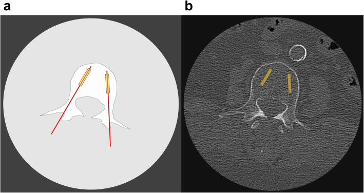
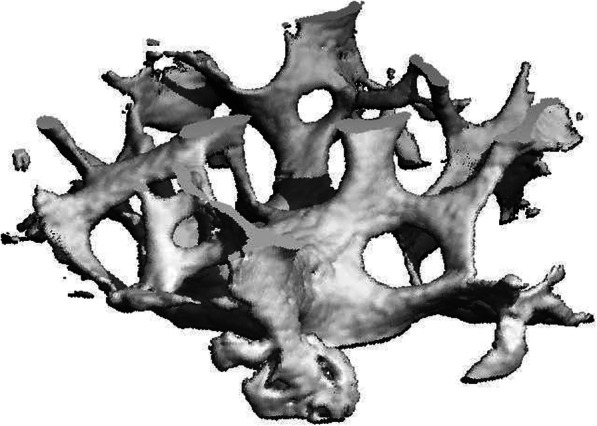
We conducted a prospective cross-sectional study to compare the results of preoperative lumbar HR-MDCT scans with those from microcomputed tomography (μCT) analysis of transpedicular vertebral body biopsies. For this purpose, we included patients undergoing spinal surgery in our orthopedic department. Each patient underwent preoperative HR-MDCT scanning (L1-L4). Intraoperatively, transpedicular biopsies were obtained from intact vertebrae. Micro-CT analysis of these biopsies was used as a reference method to assess the actual bone architecture. HR-MDCT results were statistically analyzed regarding the correlation with results from μCT.
Thirty-four patients with a mean age of 69.09 years (± 10.07) were included in the study. There was no significant correlation for any of the parameters (bone volume/total volume, trabecular separation, trabecular thickness) between μCT and HR-MDCT (bone volume/total volume: r = - 0.026 and p = 0.872; trabecular thickness: r = 0.074 and r = 6.42; and trabecular separation: r = - 0.18 and p = 0.254).
To our knowledge, this is the first study comparing in vivo HR-MDCT with μCT analysis of vertebral biopsies in human patients. Our findings suggest that lumbar HR-MDCT is not valid for the in vivo evaluation of bone architecture in the lumbar spine. New diagnostic tools for the evaluation of osteoporosis and preoperative orthopedic planning are urgently needed.
骨质疏松症的特征是骨结构和数量的恶化,导致骨折风险增加。目前,评估骨质量的主要诊断工具是双能 X 射线吸收法(DXA),但它只能测量骨量。高分辨率多探测器计算机断层扫描(HR-MDCT)提供了一种评估骨结构的替代方法,但在体内仍缺乏有效性的证据。本研究的目的是评估 HR-MDCT 评估腰椎骨结构的有效性。
我们进行了一项前瞻性的横断面研究,比较了术前腰椎 HR-MDCT 扫描结果与经椎弓根椎体活检的微计算机断层扫描(μCT)分析结果。为此,我们纳入了我院骨科行脊柱手术的患者。每位患者均接受术前腰椎 HR-MDCT 扫描(L1-L4)。术中从完整的椎骨获得经椎弓根活检。这些活检的微 CT 分析被用作评估实际骨结构的参考方法。对 HR-MDCT 结果进行统计学分析,以评估与 μCT 结果的相关性。
本研究共纳入 34 例平均年龄 69.09 岁(±10.07)的患者。μCT 和 HR-MDCT 之间的任何参数(骨体积/总体积、骨小梁分离度、骨小梁厚度)均无显著相关性(骨体积/总体积:r=-0.026,p=0.872;骨小梁厚度:r=0.074,p=0.642;骨小梁分离度:r=-0.18,p=0.254)。
据我们所知,这是首次比较体内 HR-MDCT 与人类患者椎骨活检的 μCT 分析的研究。我们的研究结果表明,腰椎 HR-MDCT 不能用于评估腰椎的骨结构。迫切需要新的诊断工具来评估骨质疏松症和术前骨科规划。