• 文献检索
  • 文档翻译
  • 深度研究
  • 学术资讯
  • Suppr Zotero 插件Zotero 插件
  • 邀请有礼
  • 套餐&价格
  • 历史记录
应用&插件
Suppr Zotero 插件Zotero 插件浏览器插件Mac 客户端Windows 客户端微信小程序
定价
高级版会员购买积分包购买API积分包
服务
文献检索文档翻译深度研究API 文档MCP 服务
关于我们
关于 Suppr公司介绍联系我们用户协议隐私条款
关注我们

Suppr 超能文献

核心技术专利:CN118964589B侵权必究
粤ICP备2023148730 号-1Suppr @ 2026

文献检索

告别复杂PubMed语法,用中文像聊天一样搜索,搜遍4000万医学文献。AI智能推荐,让科研检索更轻松。

立即免费搜索

文件翻译

保留排版,准确专业,支持PDF/Word/PPT等文件格式,支持 12+语言互译。

免费翻译文档

深度研究

AI帮你快速写综述,25分钟生成高质量综述,智能提取关键信息,辅助科研写作。

立即免费体验

通过免疫金和β2微球蛋白-金电子显微镜对变形链球菌非纤维状表面结构上β2微球蛋白(β2m)结合位点进行标记

Labeling of binding sites for beta 2-microglobulin (beta 2m) on nonfibrillar surface structures of mutans streptococci by immunogold and beta 2m-gold electron microscopy.

作者信息

Ericson D, Ellen R P, Buivids I

出版信息

J Bacteriol. 1987 Jun;169(6):2507-15. doi: 10.1128/jb.169.6.2507-2515.1987.

DOI:10.1128/jb.169.6.2507-2515.1987
PMID:3294795
原文链接:https://pmc.ncbi.nlm.nih.gov/articles/PMC212106/
Abstract

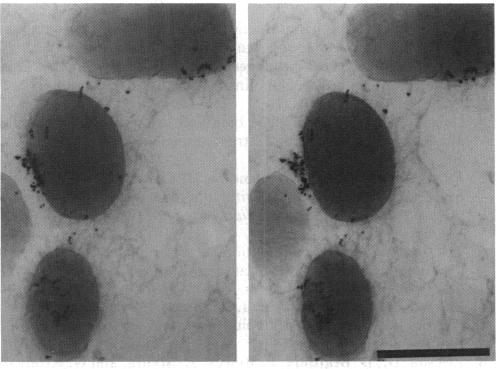
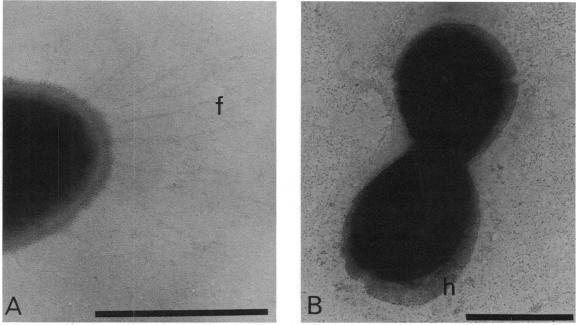
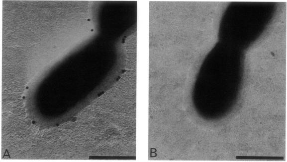
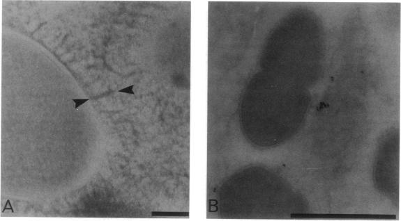
As little detail is known about the surface structure of streptococci in the mutans group and the relationship of surface structure to host ligand-binding functions, the twofold purpose of this investigation was to examine in detail, by a range of electron microscopic techniques, the surface structures of streptococci in the different species of the mutans group and to investigate the distribution of beta 2-microglobulin (beta 2m)-binding sites on such structures. Strains representing Streptococcus mutans, S. cricetus, S. rattus, S. sobrinus, and four fresh isolates were studied by shadowcasting and histochemical staining of whole-mounted cells as well as by ultrathin and thick sectioning of embedded specimens. beta 2m-binding site distribution was visualized by indirect immunogold electron microscopy and by direct bacterial binding of beta 2m-conjugated gold probes. Shadowcast preparations revealed binding of gold probes to the cell surface of known beta 2m-binding strains but not to their polar fibrillar appendages. These long fibrils, common to all strains, were trypsin and sonication sensitive and stained with lead citrate but not with uranyl acetate or ruthenium red. More gold particles were bound by the indirect technique. For grid-mounted bacteria, the gold was mostly bound in clusters at the periphery of the cells. When gold probes were reacted in suspension with bacteria before mounting onto grids, a more even distribution of the gold was seen, but the bacteria were aggregated. Heating the bacteria eliminated beta 2m-gold binding but had no effect on the morphology of the fibrils. Thick sections of embedded bacteria prereacted with beta 2m-conjugated gold probes were analyzed by stereo imaging. A wispy, uranyl acetate-stained fuzzy layer, distinct from the fibrils seen by shadowcasting and extending up to one cell diameter from the cell wall, contained the gold probes. These findings introduce a concept that binding sites for some salivary ligands on mutans streptococci may be clustered on very delicate, nonfibrillar structures extending much further from the cell wall than previously appreciated. As for beta 2m, which composes part of the human histocompatibility antigens, part of the bacterial surface would be coated at a distance from its body with a protein not necessarily recognized as foreign by the host.

摘要

由于对变形链球菌组中链球菌的表面结构以及表面结构与宿主配体结合功能之间的关系了解甚少,本研究的双重目的是通过一系列电子显微镜技术详细检查变形链球菌组不同物种中链球菌的表面结构,并研究β2-微球蛋白(β2m)结合位点在这些结构上的分布。通过对整装细胞进行投影和组织化学染色以及对包埋标本进行超薄和厚切片,对代表变形链球菌、仓鼠链球菌、大鼠链球菌、远缘链球菌的菌株以及四个新分离株进行了研究。通过间接免疫金电子显微镜和β2m偶联金探针的直接细菌结合来观察β2m结合位点的分布。投影制备显示金探针与已知β2m结合菌株的细胞表面结合,但不与它们的极性纤维状附属物结合。这些所有菌株共有的长纤维对胰蛋白酶和超声处理敏感,用柠檬酸铅染色,但不用醋酸铀或钌红染色。间接技术结合的金颗粒更多。对于网格上的细菌,金大多聚集在细胞周边。当金探针在悬浮液中与细菌反应后再固定到网格上时,金的分布更均匀,但细菌会聚集。加热细菌消除了β2m-金的结合,但对纤维的形态没有影响。通过立体成像分析了预先用β2m偶联金探针反应的包埋细菌的厚切片。一个纤细的、用醋酸铀染色的模糊层,与投影所见的纤维不同,从细胞壁延伸到一个细胞直径的距离,包含金探针。这些发现引出了一个概念,即变形链球菌上一些唾液配体的结合位点可能聚集在非常精细的、非纤维状的结构上,这些结构从细胞壁延伸的距离比以前认为的要远得多。至于构成人类组织相容性抗原一部分的β2m,细菌表面的一部分会在远离菌体的一定距离处被一种宿主不一定视为外来的蛋白质覆盖。

https://cdn.ncbi.nlm.nih.gov/pmc/blobs/566b/212106/fa373949fdcd/jbacter00196-0201-a.jpg
https://cdn.ncbi.nlm.nih.gov/pmc/blobs/566b/212106/63426186f527/jbacter00196-0197-a.jpg
https://cdn.ncbi.nlm.nih.gov/pmc/blobs/566b/212106/7377e8d8e2bb/jbacter00196-0197-b.jpg
https://cdn.ncbi.nlm.nih.gov/pmc/blobs/566b/212106/f9aa0298b9a0/jbacter00196-0198-a.jpg
https://cdn.ncbi.nlm.nih.gov/pmc/blobs/566b/212106/91617371201b/jbacter00196-0198-b.jpg
https://cdn.ncbi.nlm.nih.gov/pmc/blobs/566b/212106/b62c75e70f38/jbacter00196-0199-a.jpg
https://cdn.ncbi.nlm.nih.gov/pmc/blobs/566b/212106/dea052a2520a/jbacter00196-0199-b.jpg
https://cdn.ncbi.nlm.nih.gov/pmc/blobs/566b/212106/06d200f5e31d/jbacter00196-0200-a.jpg
https://cdn.ncbi.nlm.nih.gov/pmc/blobs/566b/212106/ef494f621c03/jbacter00196-0200-b.jpg
https://cdn.ncbi.nlm.nih.gov/pmc/blobs/566b/212106/fa373949fdcd/jbacter00196-0201-a.jpg

相似文献

1
Labeling of binding sites for beta 2-microglobulin (beta 2m) on nonfibrillar surface structures of mutans streptococci by immunogold and beta 2m-gold electron microscopy.通过免疫金和β2微球蛋白-金电子显微镜对变形链球菌非纤维状表面结构上β2微球蛋白(β2m)结合位点进行标记
J Bacteriol. 1987 Jun;169(6):2507-15. doi: 10.1128/jb.169.6.2507-2515.1987.
2
Electron microscopic localization of receptors for aggregated beta 2-microglobulin on the surface of beta-hemolytic streptococci.β-溶血性链球菌表面聚集的β2-微球蛋白受体的电子显微镜定位
Infect Immun. 1983 Oct;42(1):326-32. doi: 10.1128/iai.42.1.326-332.1983.
3
On the interaction between beta 2-microglobulin and group A streptococci.
Scand J Immunol. 1984 Jul;20(1):69-79. doi: 10.1111/j.1365-3083.1984.tb00979.x.
4
Agglutination of Streptococcus mutans by low-molecular-weight salivary components: effect of beta 2-microglobulin.低分子量唾液成分对变形链球菌的凝集作用:β2-微球蛋白的影响
Infect Immun. 1984 Nov;46(2):526-30. doi: 10.1128/iai.46.2.526-530.1984.
5
Isolation and characterization of monoclonal antibodies specific for antigen P1, a major surface protein of mutans streptococci.变形链球菌主要表面蛋白抗原P1特异性单克隆抗体的分离与鉴定
Infect Immun. 1987 Nov;55(11):2759-67. doi: 10.1128/iai.55.11.2759-2767.1987.
6
Further characteristics of beta2-microglobulin binding to oral streptococci.β2-微球蛋白与口腔链球菌结合的进一步特征
Infect Immun. 1980 Oct;30(1):117-24. doi: 10.1128/iai.30.1.117-124.1980.
7
Interactions between human serum proteins and oral streptococci reveal occurrence of receptors for aggregated beta 2-microglobulin.人血清蛋白与口腔链球菌之间的相互作用揭示了聚集的β2-微球蛋白受体的存在。
Infect Immun. 1979 Jul;25(1):279-83. doi: 10.1128/iai.25.1.279-283.1979.
8
Related bindings of aggregated beta 2-microglobulin, IgG Fab, kappa and lambda light chains to group A streptococci.聚集的β2-微球蛋白、IgG Fab、κ和λ轻链与A组链球菌的相关结合。
Acta Pathol Microbiol Immunol Scand B. 1986 Feb;94(1):25-31. doi: 10.1111/j.1699-0463.1986.tb03016.x.
9
Characterization of the beta 2-microglobulin endocytic pathway in rat proximal tubule cells.大鼠近端肾小管细胞中β2-微球蛋白内吞途径的特征
Am J Physiol. 1994 Sep;267(3 Pt 2):F380-9. doi: 10.1152/ajprenal.1994.267.3.F380.
10
Specific binding sites for monomeric and aggregated beta 2-microglobulin on surface of groups A, B, C, and G streptococci.A、B、C和G组链球菌表面上单体和聚集的β2-微球蛋白的特异性结合位点。
Microbiol Immunol. 1986;30(2):155-64. doi: 10.1111/j.1348-0421.1986.tb00930.x.

引用本文的文献

1
Saliva-binding region of Streptococcus mutans surface protein antigen.变形链球菌表面蛋白抗原的唾液结合区域
Infect Immun. 1993 Oct;61(10):4344-9. doi: 10.1128/iai.61.10.4344-4349.1993.
2
Clustering of fibronectin adhesins toward Treponema denticola tips upon contact with immobilized fibronectin.与固定化纤连蛋白接触时,纤连蛋白黏附素向齿垢密螺旋体菌毛尖端聚集。
Infect Immun. 1994 Jun;62(6):2214-21. doi: 10.1128/iai.62.6.2214-2221.1994.

本文引用的文献

1
Ultrastructure of Mutants of Streptococcus mutans with Reference to Agglutination, Adhesion, and Extracellular Polysaccharide.变形链球菌突变株的超微结构与凝集、黏附及细胞外多糖的关系
Infect Immun. 1974 Nov;10(5):1170-9. doi: 10.1128/iai.10.5.1170-1179.1974.
2
Role of salivary mucins in the protection of the oral cavity.唾液黏蛋白在口腔保护中的作用。
J Oral Pathol. 1982 Feb;11(1):1-17. doi: 10.1111/j.1600-0714.1982.tb00138.x.
3
The binding of human salivary alpha-amylase by oral strains of streptococcal bacteria.口腔链球菌菌株与人唾液α淀粉酶的结合
Arch Oral Biol. 1983;28(7):567-73. doi: 10.1016/0003-9969(83)90003-1.
4
Adherent interactions which may affect microbial ecology in the mouth.可能影响口腔微生物生态的黏附相互作用。
J Dent Res. 1984 Mar;63(3):378-85. doi: 10.1177/00220345840630030401.
5
Ability of various oral bacteria to bind human plasma fibronectin.多种口腔细菌与人血浆纤维连接蛋白结合的能力。
Microbiol Immunol. 1984;28(8):863-71. doi: 10.1111/j.1348-0421.1984.tb00742.x.
6
Agglutination of Streptococcus mutans by low-molecular-weight salivary components: effect of beta 2-microglobulin.低分子量唾液成分对变形链球菌的凝集作用:β2-微球蛋白的影响
Infect Immun. 1984 Nov;46(2):526-30. doi: 10.1128/iai.46.2.526-530.1984.
7
On the interaction between beta 2-microglobulin and group A streptococci.
Scand J Immunol. 1984 Jul;20(1):69-79. doi: 10.1111/j.1365-3083.1984.tb00979.x.
8
Electron microscopic localization of receptors for aggregated beta 2-microglobulin on the surface of beta-hemolytic streptococci.β-溶血性链球菌表面聚集的β2-微球蛋白受体的电子显微镜定位
Infect Immun. 1983 Oct;42(1):326-32. doi: 10.1128/iai.42.1.326-332.1983.
9
Streptococcus salivarius strains carry either fibrils or fimbriae on the cell surface.唾液链球菌菌株在细胞表面带有纤丝或菌毛。
J Bacteriol. 1984 Jan;157(1):64-72. doi: 10.1128/jb.157.1.64-72.1984.
10
Further characteristics of beta2-microglobulin binding to oral streptococci.β2-微球蛋白与口腔链球菌结合的进一步特征
Infect Immun. 1980 Oct;30(1):117-24. doi: 10.1128/iai.30.1.117-124.1980.