Tigabie Workye, Tesfay Hadush, Tamrat Dagnachew, Raya Kassahun, Negussie Tihitena
Addis Ababa University, College of Health Sciences, Ethiopia.
Int J Surg Case Rep. 2020;75:117-121. doi: 10.1016/j.ijscr.2020.09.023. Epub 2020 Sep 11.
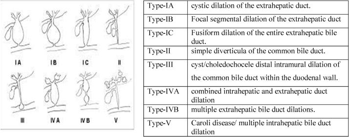
Choledochal cyst (CC) is an uncommon congenital disease of the biliary tract. There are five main types of CC with several recognized sub-types. However, occasional variants with a difficulty in diagnosis and management do occur.
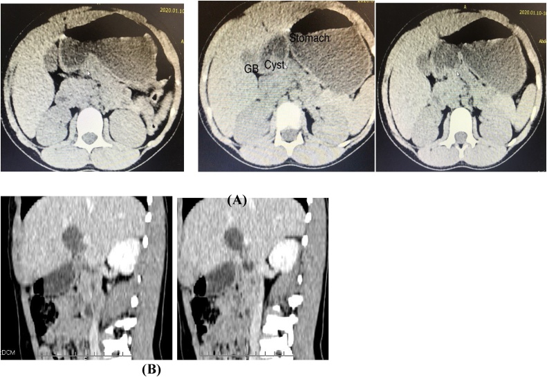
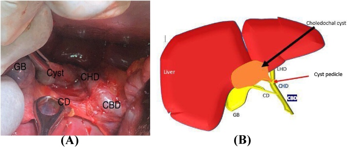
We report a case of a nine years old female child diagnosed with CC who presented with right quadrant abdominal pain with unremarkable physical findings. Investigation using abdominal CT scan suggested type II choledochal cyst. The intraoperative finding revealed an unusual site of the cyst that is at the confluence of common hepatic duct (CHD) posteriorly. The cyst was successfully excised and the child is doing well on her follow ups.
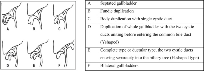
In the management of choledochal cyst the anatomy should be clearly defined with detailed investigations like Abdominal CT Scan or cholangiography before surgical excision as abnormal variants which usually do not fit into the known classification types and subtypes. This might confuse with other differentials like gall bladder duplication. Surgical excision is the gold standard management option.
This case report will alert surgeons that there are different anatomic variant of choledochal cysts out of the known classifications and with meticulous dissection will help proper excision and avoid unnecessary complications.
胆总管囊肿(CC)是一种罕见的先天性胆道疾病。CC主要有五种类型,还有几种公认的亚型。然而,偶尔也会出现诊断和治疗困难的变异型。
我们报告一例9岁女性儿童被诊断为CC,表现为右下腹疼痛,体格检查无异常发现。腹部CT扫描检查提示为II型胆总管囊肿。术中发现囊肿位于肝总管(CHD)后方汇合处这一不寻常位置。囊肿被成功切除,患儿随访情况良好。
在胆总管囊肿的治疗中,手术切除前应通过腹部CT扫描或胆管造影等详细检查明确解剖结构,因为异常变异型通常不符合已知的分类类型和亚型。这可能会与胆囊重复等其他鉴别诊断相混淆。手术切除是金标准治疗选择。
本病例报告将提醒外科医生,胆总管囊肿存在已知分类之外的不同解剖变异型,细致的解剖有助于正确切除并避免不必要的并发症。