Department of Pathology and Biological Responses, Nagoya University Graduate School of Medicine, 65 Tsurumai-cho, Showa-Ku, Nagoya, 466-8550, Japan; Department of Obstetrics and Gynecology, Nagoya University Graduate School of Medicine, 65 Tsurumai-cho, Showa-Ku, Nagoya, 466-8550, Japan.
Department of Obstetrics and Gynecology, Nagoya University Graduate School of Medicine, 65 Tsurumai-cho, Showa-Ku, Nagoya, 466-8550, Japan.
Redox Biol. 2020 Oct;37:101726. doi: 10.1016/j.redox.2020.101726. Epub 2020 Sep 15.
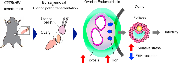
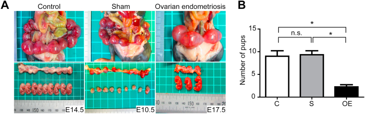
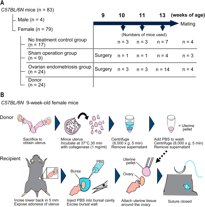
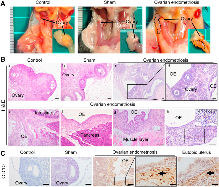
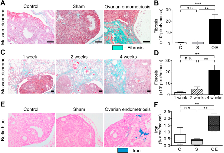
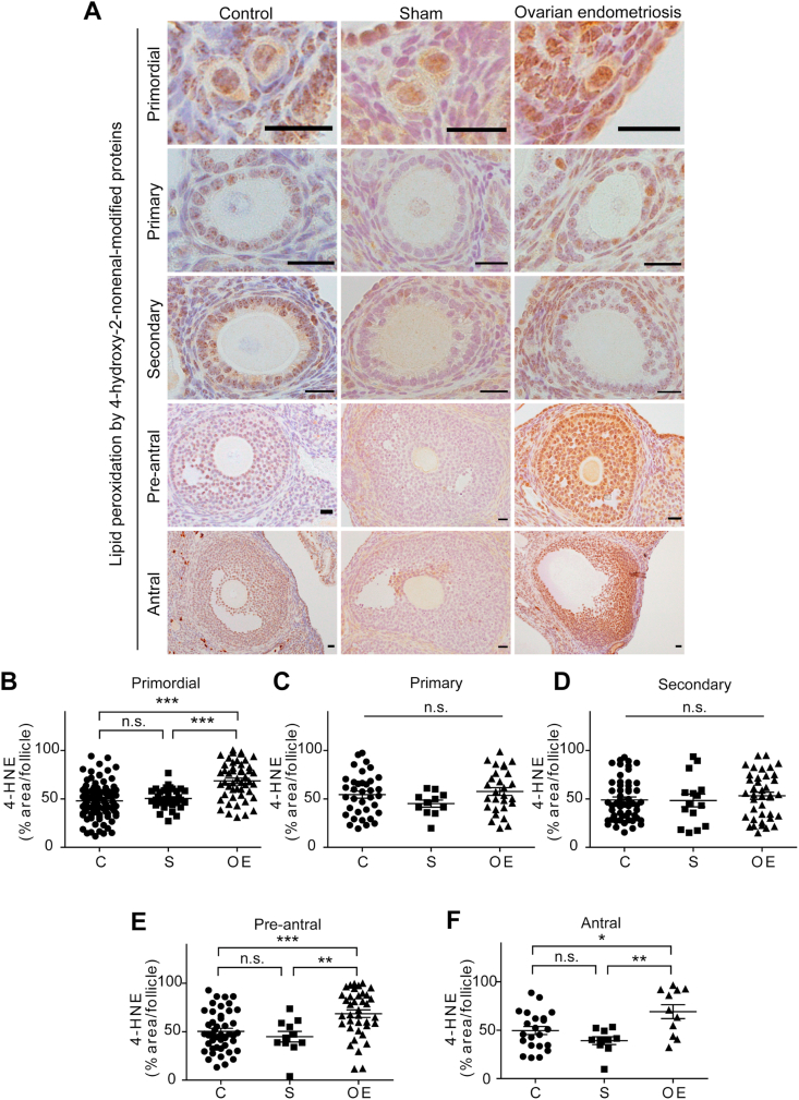
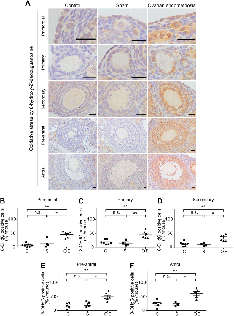
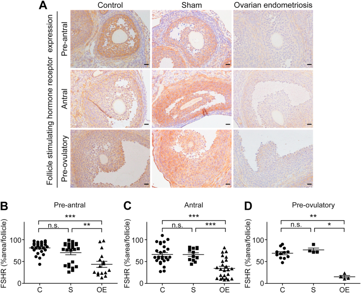
Ovarian endometriosis (OE) provides women of reproductive age with not only severe menstrual pain but also infertility and an increased risk for ovarian carcinogenesis. Whereas peritoneal endometriosis models have been developed with syngeneic implantation of minced uterine tissue and oncogenic K-ras allele with conditional Pten deletion within ovarian surface epithelium generated preneoplastic endometrial glandular morphology, followed by endometrioid adenocarcinoma, there has been no mouse model of OE similar to human counterparts, applicable to preclinical studies. Here we for the first time established a murine OE model that reveals infertility, and evaluated the involvement of iron catalyzed oxidative stress in the pathogenesis. Minced uterine tissue from female mice was implanted on ovarian surface of syngeneic mice after bursectomy to induce OE. Ectopic growth of endometrium was observed in association with ovary 4 weeks after implantation in 85.7% (12/14) of the operated mice with our protocol. Endometriotic lesions involved intestine, pancreas and peritoneal wall. Fibrosis around the ovary was prominent and increased time-dependently in the OE group. Iron accumulation was significantly increased in the OE group, leading to oxidative stress in each stage of the follicles as evaluated by 4-hydroxy-2-nonenal-modified proteins and 8-hydroxy-2'-deoxyguanosine. Expression of follicle stimulating hormone receptor in the follicles revealed a significant decrease during pre-antral, antral and pre-ovulatory phases in the OE group. Finally, the number of pups was significantly reduced in the OE group in comparison to the controls. This model affords an opportunity to evaluate agents or procedures to counteract ovarian endometriosis in the preclinical settings.
卵巢子宫内膜异位症 (OE) 不仅给育龄妇女带来严重的月经疼痛,还导致不孕和卵巢癌发生风险增加。虽然已经建立了腹膜子宫内膜异位症模型,通过同种异体植入小块子宫组织和卵巢表面上皮中的致癌 K-ras 等位基因与条件性 Pten 缺失,产生了具有preneoplastic 子宫内膜腺形态的子宫内膜异位症,随后是子宫内膜样腺癌,但还没有类似于人类的 OE 小鼠模型,适用于临床前研究。在这里,我们首次建立了一种小鼠 OE 模型,该模型揭示了不孕,并评估了铁催化的氧化应激在发病机制中的作用。切除囊后,将来自雌性小鼠的子宫组织小块植入同种异体小鼠的卵巢表面,以诱导 OE。在我们的方案中,85.7%(12/14)的手术小鼠在植入后 4 周观察到子宫内膜异位生长,与卵巢相关。异位性病变涉及肠、胰腺和腹膜壁。卵巢周围的纤维化在 OE 组中明显且随时间增加。OE 组中铁的积累显著增加,导致每个卵泡阶段的氧化应激,如通过 4-羟基-2-壬烯醛修饰蛋白和 8-羟基-2'-脱氧鸟苷评估。在 OE 组中,卵泡刺激素受体在卵泡中的表达在前期、腔前期和排卵前期均显著降低。最后,OE 组的幼仔数量明显少于对照组。该模型为在临床前环境中评估对抗卵巢子宫内膜异位症的药物或程序提供了机会。