Hormone Laboratory, Department of Medical Biochemistry and Pharmacology, Haukeland University Hospital, Bergen, Norway.
Department of Clinical Science, University of Bergen, Bergen, Norway.
J Clin Endocrinol Metab. 2020 Dec 1;105(12):e4886-95. doi: 10.1210/clinem/dgaa679.
Application of ultrasound (US) to evaluate attainment and morphology of glandular tissue provides a new rationale for evaluating onset and progression of female puberty, but currently no hormone references complement this method. Furthermore, previous studies have not explored the predictive value of endocrine profiling to determine female puberty onset.
To integrate US breast staging with hypothalamic-pituitary-gonadal hormone references and test the predictive value of an endocrine profile to determine thelarche.
Cross-sectional sample of 601 healthy Norwegian girls, ages 6 to 16 years.
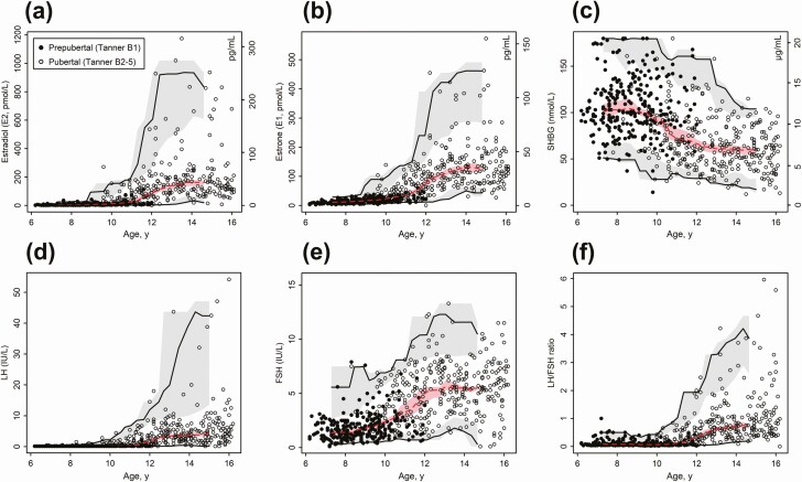
Clinical and ultrasound breast evaluations were performed for all included girls. Blood samples were analyzed by immunoassay and ultrasensitive liquid chromatography-tandem mass spectrometry (LC-MS/MS) to quantify estradiol (E2) and estrone (E1) from the subpicomolar range.
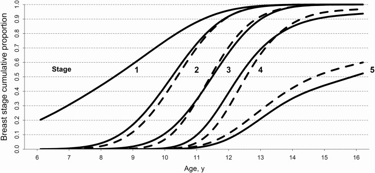
References for E2, E1, luteinizing hormone, follicle-stimulating hormone, and sex hormone-binding globulin were constructed in relation to chronological age, Tanner stages, and US breast stages. An endocrine profile index score derived from principal component analysis of these analytes was a better marker of puberty onset than age or any individual hormone, with receiver-operating characteristic area under the curve 0.91 (P < 0.001). Ultrasound detection of nonpalpable glandular tissue in 14 out of 264 (5.3%) girls with clinically prepubertal presentation was associated with significantly higher median serum levels of E2 (12.5 vs 4.9 pmol/L; P < 0.05) and a distinct endocrine profile (arbitrary units; P < 0.001).
We provide the first hormone references for use with US breast staging and demonstrate the application of endocrine profiling to improve detection of female puberty onset.
超声(US)在评估腺体组织的发育和形态方面的应用为评估女性青春期的开始和进展提供了新的依据,但目前尚无激素参考值来补充这种方法。此外,以前的研究尚未探讨内分泌谱预测值来确定女性青春期开始。
将 US 乳房分期与下丘脑-垂体-性腺激素参考值相结合,并测试内分泌谱预测值以确定乳房发育开始。
设计、地点和参与者:横断面样本为 601 名健康的挪威女孩,年龄 6 至 16 岁。
对所有纳入的女孩进行临床和超声乳房评估。通过免疫测定法和超敏液相色谱-串联质谱(LC-MS/MS)分析血液样本,以从小分子范围内定量雌二醇(E2)和雌酮(E1)。
构建了 E2、E1、促黄体生成素、促卵泡生成素和性激素结合球蛋白与实际年龄、Tanner 分期和 US 乳房分期相关的参考值。这些分析物的主成分分析得出的内分泌谱指数评分是青春期开始的更好标志物,优于年龄或任何单个激素,其接收者操作特征曲线下面积为 0.91(P < 0.001)。在 264 名临床性青春期前表现的女孩中,有 14 名女孩的超声检测到不可触及的腺体组织,其血清 E2(12.5 与 4.9 pmol/L;P < 0.05)和独特的内分泌谱(任意单位;P < 0.001)中位数显著更高。
我们提供了首个与 US 乳房分期一起使用的激素参考值,并展示了内分泌谱在提高女性青春期开始检测中的应用。