Shanbhag Siddharth, Suliman Salwa, Bolstad Anne Isine, Stavropoulos Andreas, Mustafa Kamal
Department of Clinical Dentistry, Faculty of Medicine, University of Bergen, Bergen, Norway.
Department of Periodontology, Faculty of Odontology, Malmö University, Malmö, Sweden.
Front Bioeng Biotechnol. 2020 Aug 19;8:968. doi: 10.3389/fbioe.2020.00968. eCollection 2020.
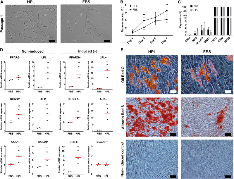
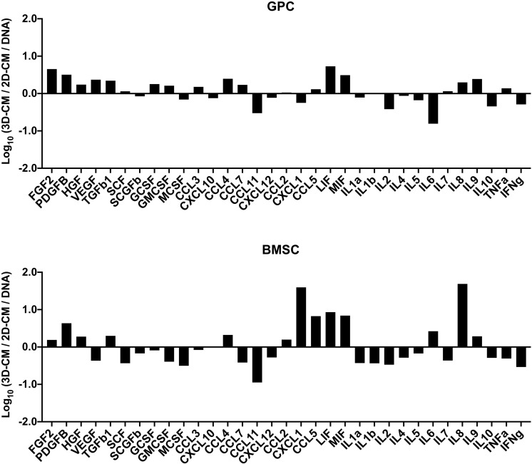
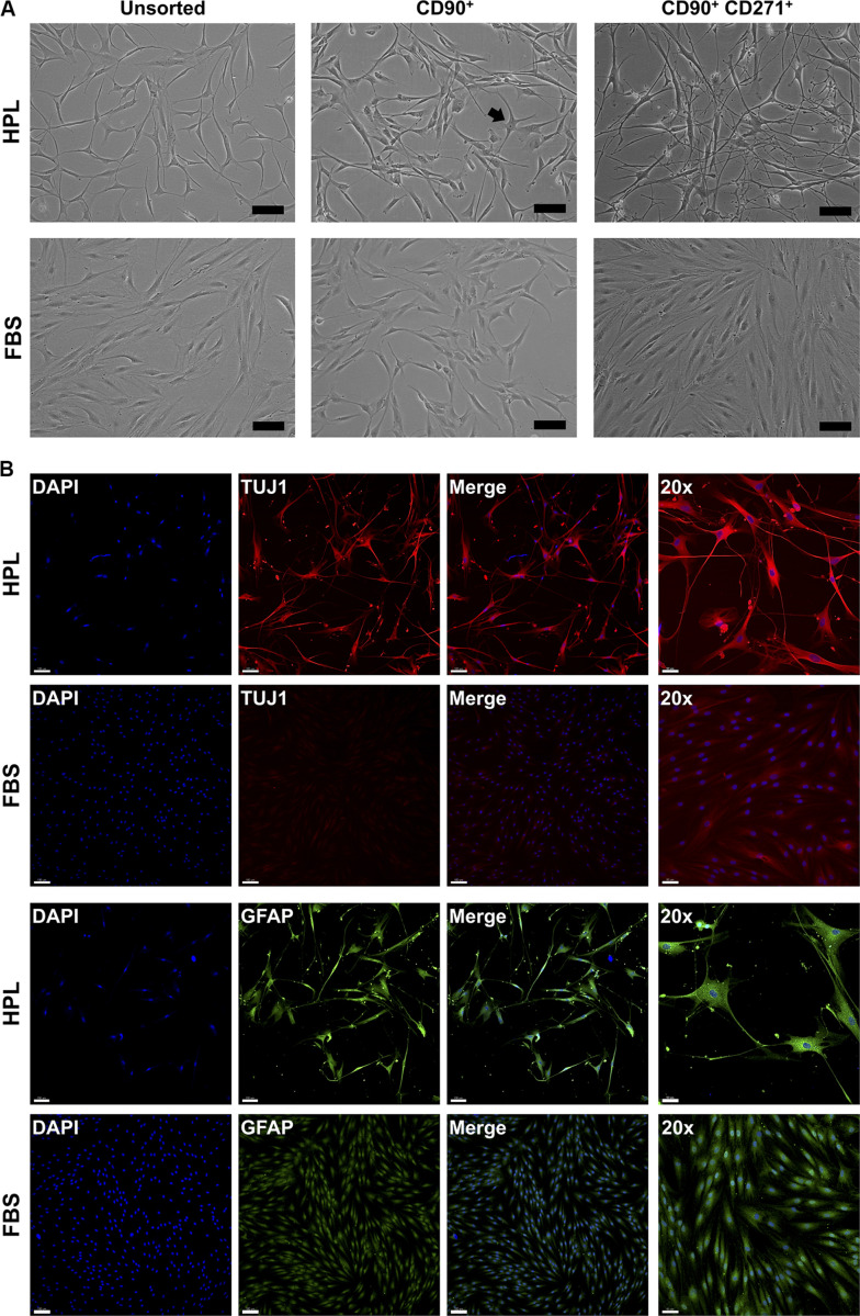
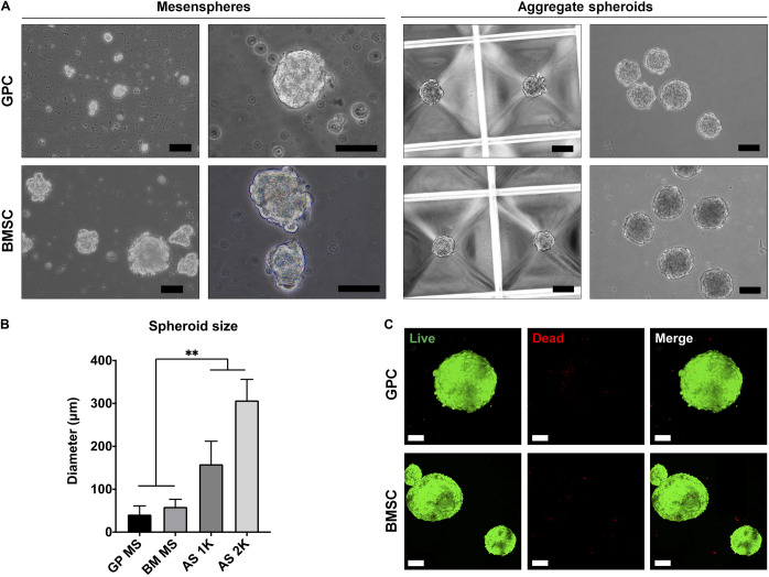
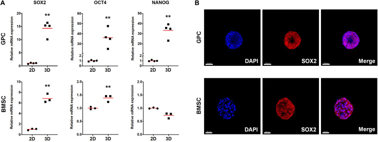
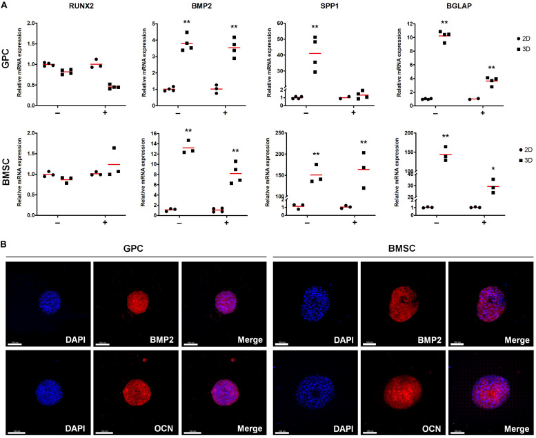
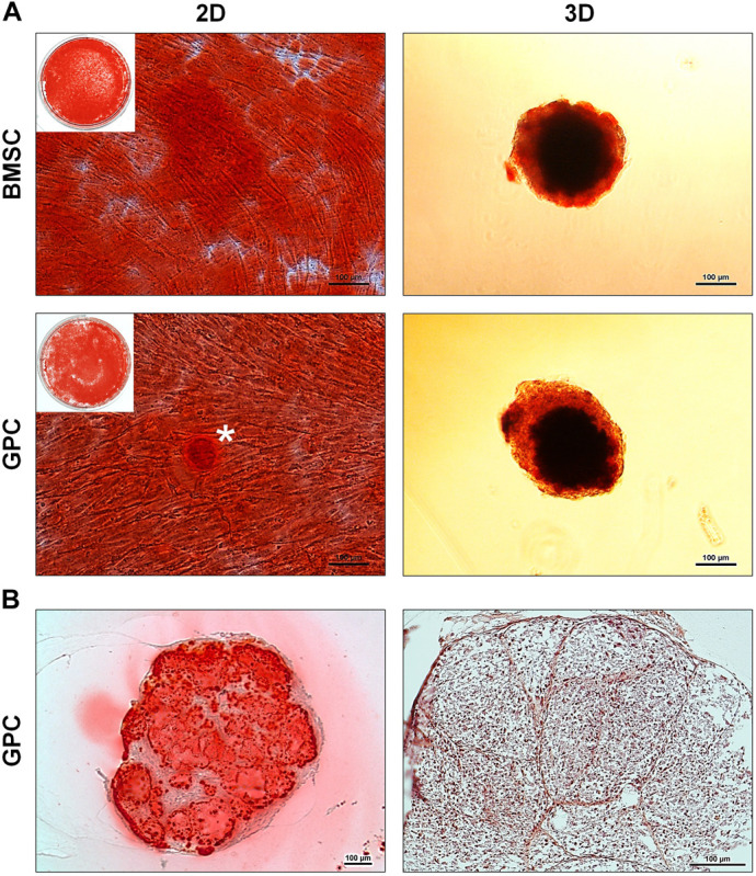
Gingiva has been identified as a minimally invasive source of multipotent progenitor cells (GPCs) for use in bone tissue engineering (BTE). To facilitate clinical translation, it is important to characterize GPCs in xeno-free cultures. Recent evidence indicates several advantages of three-dimensional (3D) spheroid cultures of mesenchymal stromal cells (MSCs) over conventional 2D monolayers. The present study aimed to characterize human GPCs in xeno-free 2D cultures, and to test their osteogenic potential in 3D cultures, in comparison to bone marrow MSCs (BMSCs). Primary GPCs and BMSCs were expanded in human platelet lysate (HPL) or fetal bovine serum (FBS) and characterized based on proliferation, immunophenotype and multi-lineage differentiation. Next, 3D spheroids of GPCs and BMSCs were formed via self-assembly and cultured in HPL. Expression of stemness- (SOX2, OCT4, NANOG) and osteogenesis-related markers (BMP2, RUNX2, OPN, OCN) was assessed at gene and protein levels in 3D and 2D cultures. The cytokine profile of 3D and 2D GPCs and BMSCs was assessed via a multiplex immunoassay. Monolayer GPCs in both HPL and FBS demonstrated a characteristic MSC-like immunophenotype and multi-lineage differentiation; osteogenic differentiation of GPCs was enhanced in HPL vs. FBS. CD271 GPCs in HPL spontaneously acquired a neuronal phenotype and strongly expressed neuronal/glial markers. 3D spheroids of GPCs and BMSCs with high cell viability were formed in HPL media. Expression of stemness- and osteogenesis-related genes was significantly upregulated in 3D vs. 2D GPCs/BMSCs; the latter was independent of osteogenic induction. Synthesis of SOX2, BMP2 and OCN was confirmed via immunostaining, and mineralization via Alizarin red staining. Finally, secretion of several growth factors and chemokines was enhanced in GPC/BMSC spheroids, while that of pro-inflammatory cytokines was reduced, compared to monolayers. In summary, monolayer GPCs expanded in HPL demonstrate enhanced osteogenic differentiation potential, comparable to that of BMSCs. Xeno-free spheroid culture further enhances stemness- and osteogenesis-related gene expression, and cytokine secretion in GPCs, comparable to that of BMSCs.
牙龈已被确定为用于骨组织工程(BTE)的多能祖细胞(GPC)的微创来源。为了促进临床转化,在无血清培养中对GPC进行表征很重要。最近的证据表明,间充质基质细胞(MSC)的三维(3D)球体培养比传统的二维单层培养有几个优点。本研究旨在表征无血清二维培养中的人GPC,并与骨髓间充质干细胞(BMSC)相比,测试其在三维培养中的成骨潜力。原代GPC和BMSC在人血小板裂解物(HPL)或胎牛血清(FBS)中扩增,并根据增殖、免疫表型和多向分化进行表征。接下来,GPC和BMSC的三维球体通过自组装形成,并在HPL中培养。在三维和二维培养中,在基因和蛋白质水平评估干性相关标志物(SOX2、OCT4、NANOG)和成骨相关标志物(BMP2、RUNX2、OPN、OCN)的表达。通过多重免疫测定评估三维和二维GPC和BMSC的细胞因子谱。HPL和FBS中的单层GPC均表现出典型的MSC样免疫表型和多向分化;与FBS相比,HPL中GPC的成骨分化增强。HPL中的CD271 GPC自发获得神经元表型并强烈表达神经元/神经胶质标志物。在HPL培养基中形成了具有高细胞活力的GPC和BMSC三维球体。与二维GPC/BMSC相比,三维GPC/BMSC中干性和成骨相关基因的表达显著上调;后者与成骨诱导无关。通过免疫染色证实了SOX2、BMP2和OCN的合成,并通过茜素红染色证实了矿化。最后,与单层相比,GPC/BMSC球体中几种生长因子和趋化因子的分泌增加,而促炎细胞因子的分泌减少。总之,在HPL中扩增的单层GPC表现出增强的成骨分化潜力,与BMSC相当。无血清球体培养进一步增强了GPC中干性和成骨相关基因的表达以及细胞因子分泌,与BMSC相当。