Kim Jeonghyun, Adachi Taiji
Biomechanics Laboratory, Institute for Frontier Life and Medical Sciences, Kyoto University, Kyoto, Japan.
Front Bioeng Biotechnol. 2019 Oct 23;7:288. doi: 10.3389/fbioe.2019.00288. eCollection 2019.
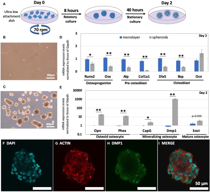
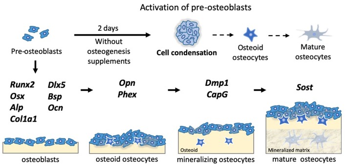
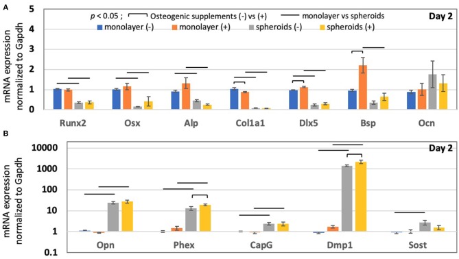
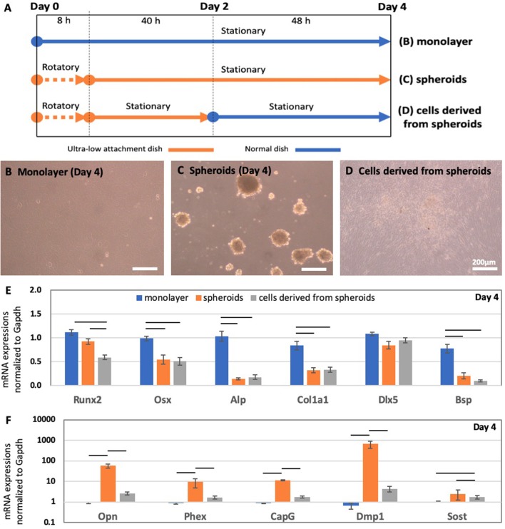
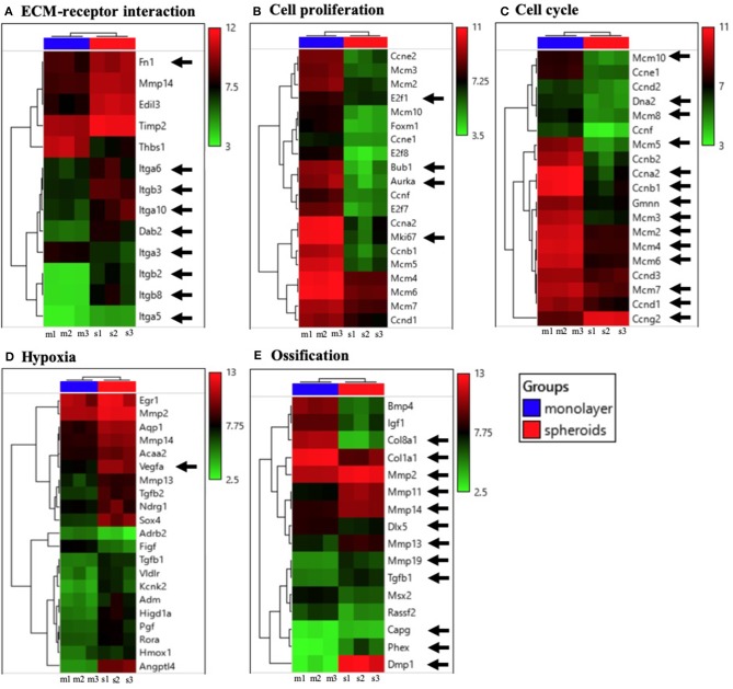
Though the three-dimensional (3D) culture system has received attention as a powerful tool for conducting biological research, bone formation and osteocyte differentiation studies have mostly been based on results obtained using two-dimensional (2D) culture systems. Here, we introduced a rotatory culture system to fabricate 3D spheroids, using mouse osteoblast precursor cells. These spheroids, incubated for 2 days without chemical induction by osteogenic supplements, exhibited notably up-regulated osteocyte marker levels; osteoblast marker levels were down-regulated, as compared to those of the conventional 2D monolayer model. The cell condensation achieved with the 3D spheroid structure triggered a greater level of differentiation of osteoblast precursor cells into osteocyte-like cells than that observed during chemical induction. Our study might imply that osteoblasts proliferate and become condensed at the targeted bone remodeling site, because of which osteoblasts achieved the capability to differentiate into osteocytes .
尽管三维(3D)培养系统作为进行生物学研究的有力工具已受到关注,但骨形成和骨细胞分化研究大多基于使用二维(2D)培养系统获得的结果。在此,我们引入了一种旋转培养系统,利用小鼠成骨细胞前体细胞制造3D球体。这些球体在没有成骨补充剂化学诱导的情况下培养2天,显示出骨细胞标志物水平显著上调;与传统的2D单层模型相比,成骨细胞标志物水平下调。与化学诱导过程中观察到的情况相比,3D球体结构实现的细胞凝聚触发了成骨细胞前体细胞向骨细胞样细胞更高水平的分化。我们的研究可能意味着成骨细胞在目标骨重塑部位增殖并凝聚,因此成骨细胞获得了分化为骨细胞的能力。