Center for Translational Oral Research (TOR), Department of Clinical Dentistry, Faculty of Medicine, University of Bergen, Årstadveien 19, 5009, Bergen, Norway.
Department of Immunology and Transfusion Medicine, Haukeland University Hospital, Bergen, Norway.
Stem Cell Res Ther. 2021 Nov 14;12(1):575. doi: 10.1186/s13287-021-02642-w.
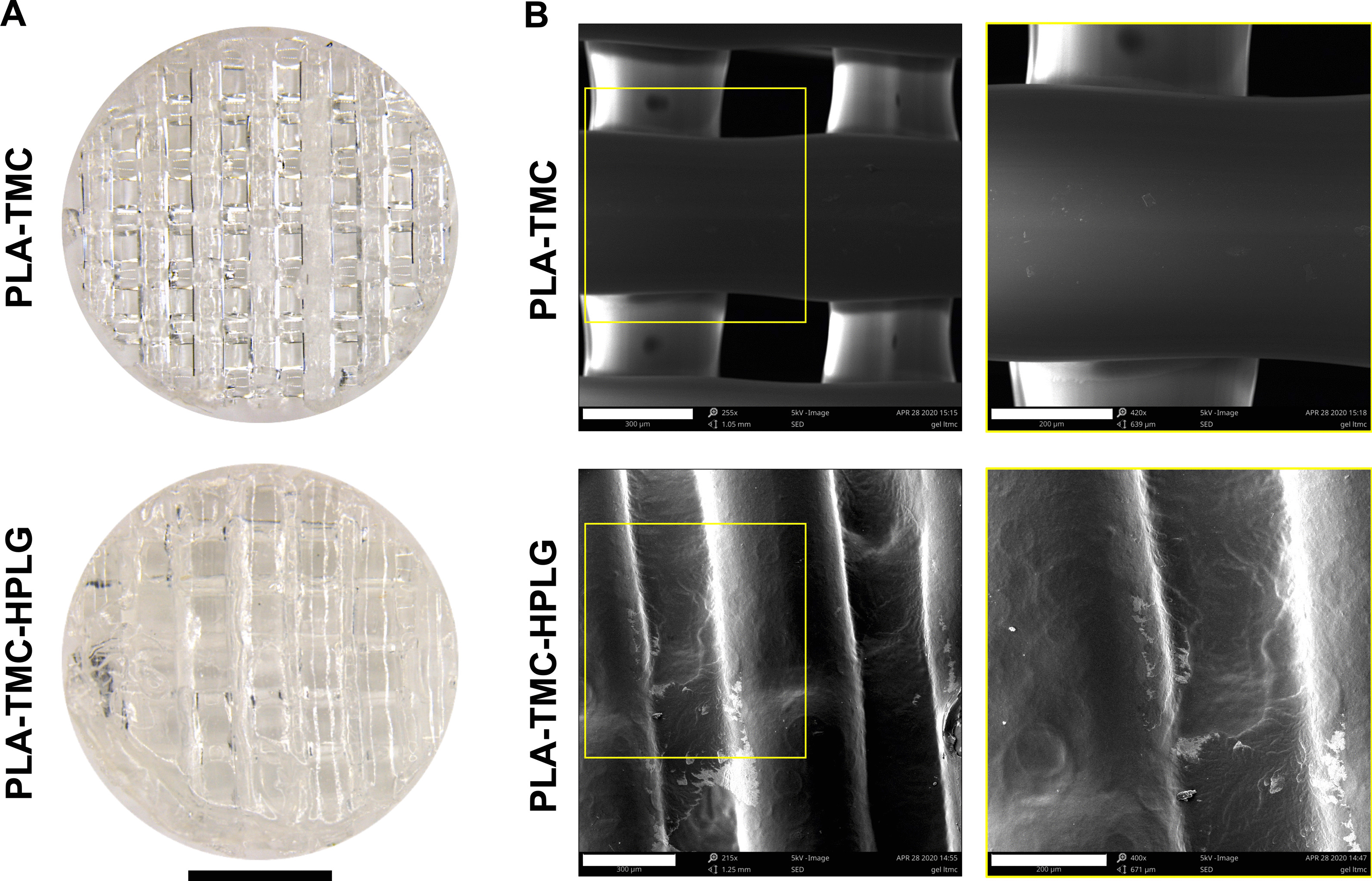
Three-dimensional (3D) spheroid culture can promote the osteogenic differentiation of bone marrow mesenchymal stromal cells (BMSC). 3D printing offers the possibility to produce customized scaffolds for complex bone defects. The aim of this study was to compare the potential of human BMSC cultured as 2D monolayers or 3D spheroids encapsulated in constructs of 3D-printed poly-L-lactide-co-trimethylene carbonate scaffolds and modified human platelet lysate hydrogels (PLATMC-HPLG) for bone regeneration.
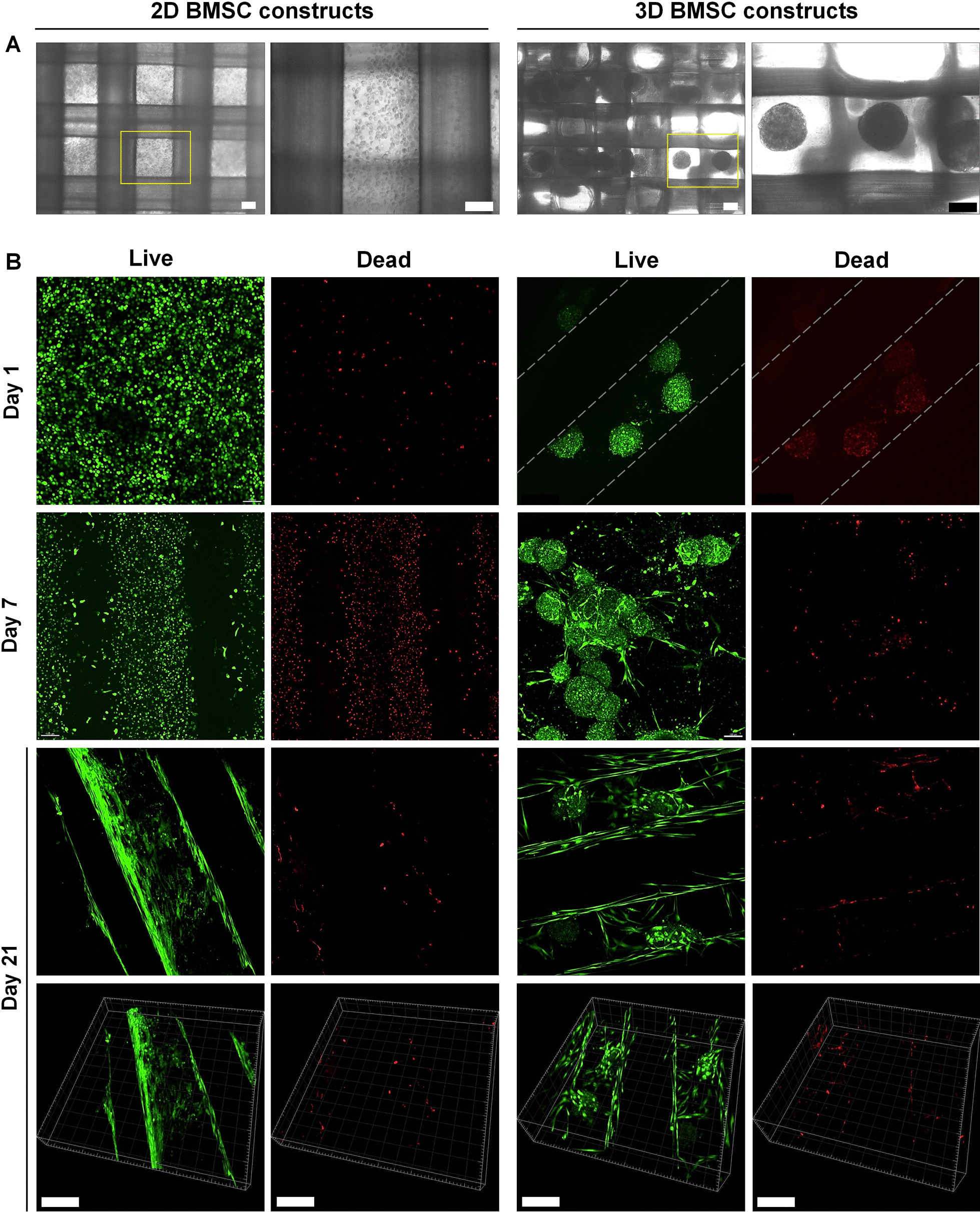
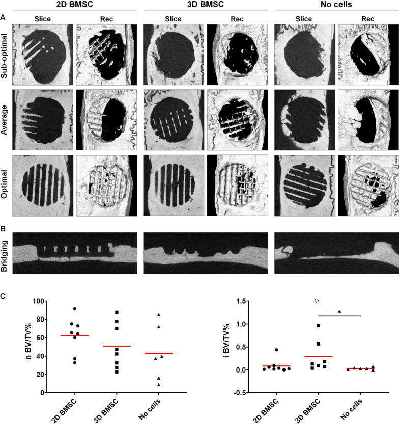
PLATMC-HPLG constructs with 2D or 3D BMSC were assessed for osteogenic differentiation based on gene expression and in vitro mineralization. Subsequently, PLATMC-HPLG constructs with 2D or 3D BMSC were implanted in rat calvarial defects for 12 weeks; cell-free constructs served as controls. Bone regeneration was assessed via in vivo computed tomography (CT), ex vivo micro-CT and histology.
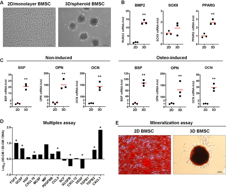
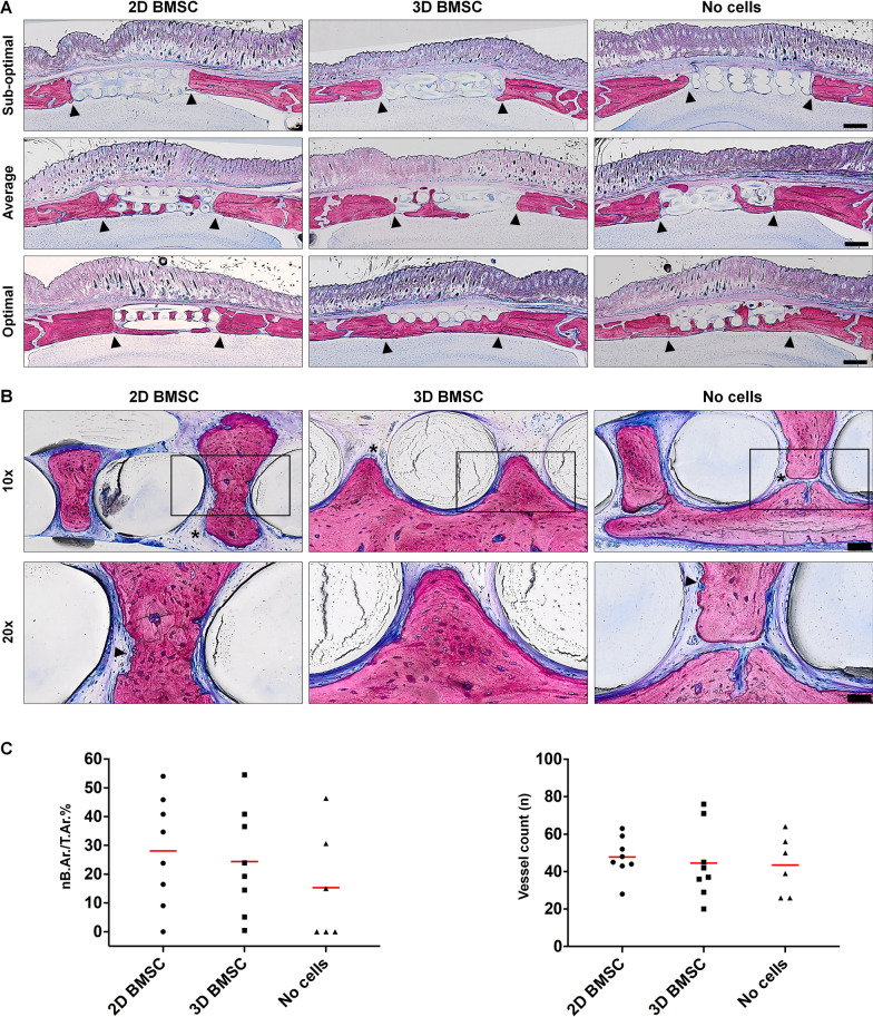
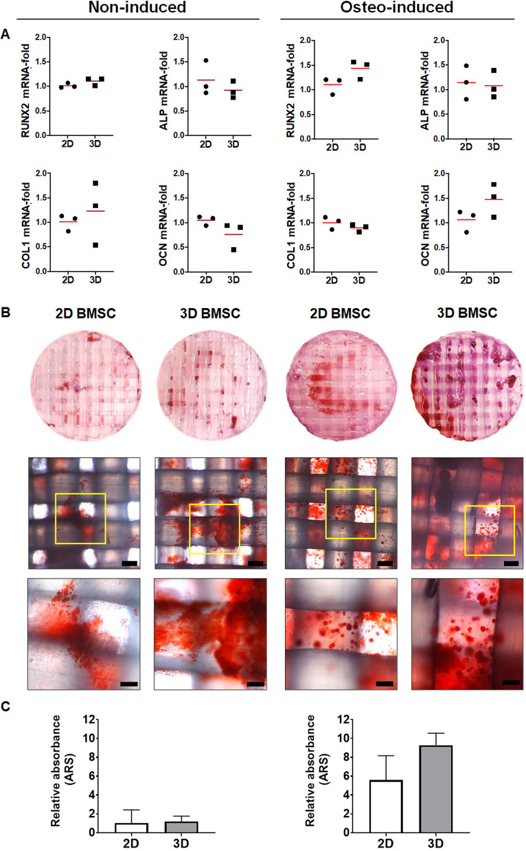
Osteogenic gene expression was significantly enhanced in 3D versus 2D BMSC prior to, but not after, encapsulation in PLATMC-HPLG constructs. A trend for greater in vitro mineralization was observed in constructs with 3D versus 2D BMSC (p > 0.05). In vivo CT revealed comparable bone formation after 4, 8 and 12 weeks in all groups. After 12 weeks, micro-CT revealed substantial regeneration in 2D BMSC (62.47 ± 19.46%), 3D BMSC (51.01 ± 24.43%) and cell-free PLATMC-HPLG constructs (43.20 ± 30.09%) (p > 0.05). A similar trend was observed in the histological analysis.
Despite a trend for superior in vitro mineralization, constructs with 3D and 2D BMSC performed similarly in vivo. Regardless of monolayer or spheroid cell culture, PLATMC-HPLG constructs represent promising scaffolds for bone tissue engineering applications.
三维(3D)球体培养可以促进骨髓间充质基质细胞(BMSC)的成骨分化。3D 打印为生产复杂骨缺损的定制支架提供了可能性。本研究的目的是比较作为 2D 单层或 3D 球体包封在 3D 打印聚-L-乳酸-共-三亚甲基碳酸酯支架和改良的人血小板裂解物水凝胶(PLATMC-HPLG)构建体中的人 BMSC 的潜力,用于骨再生。
根据基因表达和体外矿化,评估 2D 或 3D BMSC 的 PLATMC-HPLG 构建体的成骨分化。随后,将 2D 或 3D BMSC 的 PLATMC-HPLG 构建体植入大鼠颅骨缺损中 12 周;无细胞构建体作为对照。通过体内计算机断层扫描(CT)、体外 micro-CT 和组织学评估骨再生。
在封装在 PLATMC-HPLG 构建体之前,但不在之后,3D 比 2D BMSC 的成骨基因表达显著增强。在 3D 与 2D BMSC 构建体中观察到体外矿化趋势更大(p > 0.05)。体内 CT 显示所有组在 4、8 和 12 周后骨形成相似。12 周后,micro-CT 显示 2D BMSC(62.47 ± 19.46%)、3D BMSC(51.01 ± 24.43%)和无细胞 PLATMC-HPLG 构建体(43.20 ± 30.09%)有大量再生(p > 0.05)。组织学分析也观察到类似的趋势。
尽管体外矿化有趋势更好,但 3D 和 2D BMSC 的构建体在体内表现相似。无论单层还是球体细胞培养,PLATMC-HPLG 构建体都代表了用于骨组织工程应用的有前途的支架。