Wang Song, Zhang Li-Cheng, Fu Hai-Tao, Deng Jun-Hao, Xu Gao-Xiang, Li Tong, Ji Xin-Ran, Tang Pei-Fu
School of Medicine, Nankai University, Tianjin; Department of Orthopedics, Chinese PLA General Hospital, Beijing, China.
Department of Orthopedics, Chinese PLA General Hospital, Beijing, China.
Neural Regen Res. 2021 Mar;16(3):573-579. doi: 10.4103/1673-5374.290905.
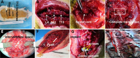
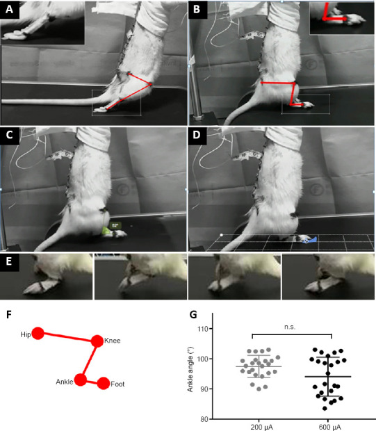
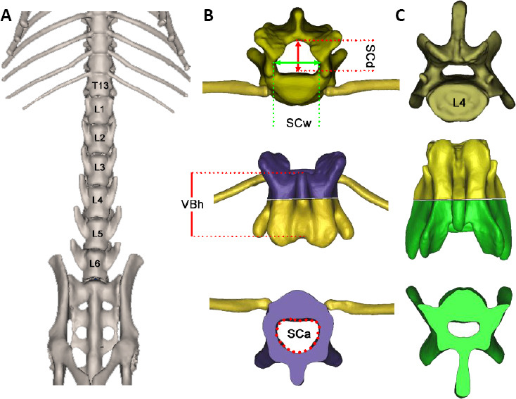
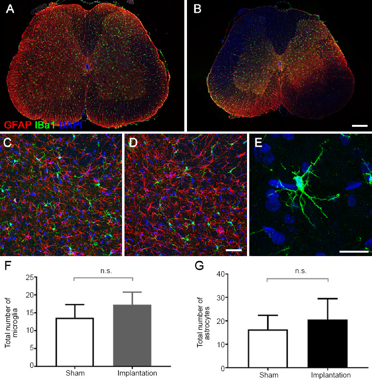
Epidural electrical stimulation can restore limb motor function after spinal cord injury by reactivating the surviving neural circuits. In previous epidural electrical stimulation studies, single electrode sites and continuous tetanic stimulation have often been used. With this stimulation, the body is prone to declines in tolerance and locomotion coordination. In the present study, rat models of complete spinal cord injury were established by vertically cutting the spinal cord at the T8 level to eliminate disturbance from residual nerve fibers, and were then subjected to epidural electrical stimulation. The flexible extradural electrode had good anatomical topology and matched the shape of the spinal canal of the implanted segment. Simultaneously, the electrode stimulation site was able to be accurately applied to the L2-3 and S1 segments of the spinal cord. To evaluate the biocompatibility of the implanted epidural electrical stimulation electrodes, GFAP/Iba-1 double-labeled immunofluorescence staining was performed on the spinal cord below the electrodes at 7 days after the electrode implantation. Immunofluorescence results revealed no significant differences in the numbers or morphologies of microglia and astrocytes in the spinal cord after electrode implantation, and there was no activated Iba-1 cell aggregation, indicating that the implant did not cause an inflammatory response in the spinal cord. Rat gait analysis showed that, at 3 days after surgery, gait became coordinated in rats with spinal cord injury under burst stimulation. The regained locomotion could clearly distinguish the support phase and the swing phase and dynamically adjust with the frequency of stimulus distribution. To evaluate the matching degree between the flexible epidural electrode (including three stimulation contacts), vertebral morphology, and the level of the epidural site of the stimulation electrode, micro-CT was used to scan the thoracolumbar vertebrae of rats before and after electrode implantation. Based on the experimental results of gait recovery using three-site stimulation electrodes at L2-3 and S1 combined with burst stimulation in a rat model of spinal cord injury, epidural electrical stimulation is a promising protocol that needs to be further explored. This study was approved by the Animal Ethics Committee of Chinese PLA General Hospital (approval No. 2019-X15-39) on April 19, 2019.
硬膜外电刺激可通过重新激活存活的神经回路来恢复脊髓损伤后的肢体运动功能。在以往的硬膜外电刺激研究中,常使用单电极位点和连续强直刺激。采用这种刺激方式时,身体容易出现耐受性下降和运动协调性降低的情况。在本研究中,通过在T8水平垂直切断脊髓建立完全性脊髓损伤大鼠模型,以消除残留神经纤维的干扰,然后对其进行硬膜外电刺激。柔性硬膜外电极具有良好的解剖拓扑结构,与植入节段的椎管形状相匹配。同时,电极刺激位点能够准确地施加于脊髓的L2-3和S1节段。为评估植入的硬膜外电刺激电极的生物相容性,在电极植入后7天,对电极下方的脊髓进行GFAP/Iba-1双标免疫荧光染色。免疫荧光结果显示,电极植入后脊髓中小胶质细胞和星形胶质细胞的数量及形态无显著差异,且无活化的Iba-1细胞聚集,表明植入物未在脊髓中引起炎症反应。大鼠步态分析表明,术后3天,脊髓损伤大鼠在爆发性刺激下步态变得协调。恢复的运动能够清晰区分支撑期和摆动期,并随刺激分布频率动态调整。为评估柔性硬膜外电极(包括三个刺激触点)、椎体形态与刺激电极硬膜外位点水平之间的匹配程度,在电极植入前后使用微型CT扫描大鼠的胸腰椎。基于在脊髓损伤大鼠模型中使用L2-3和S1的三点刺激电极联合爆发性刺激进行步态恢复的实验结果,硬膜外电刺激是一种有前景的方案,有待进一步探索。本研究于2019年4月19日获得中国人民解放军总医院动物伦理委员会批准(批准号:2019-X15-39)。