• 文献检索
  • 文档翻译
  • 深度研究
  • 学术资讯
  • Suppr Zotero 插件Zotero 插件
  • 邀请有礼
  • 套餐&价格
  • 历史记录
应用&插件
Suppr Zotero 插件Zotero 插件浏览器插件Mac 客户端Windows 客户端微信小程序
定价
高级版会员购买积分包购买API积分包
服务
文献检索文档翻译深度研究API 文档MCP 服务
关于我们
关于 Suppr公司介绍联系我们用户协议隐私条款
关注我们

Suppr 超能文献

核心技术专利:CN118964589B侵权必究
粤ICP备2023148730 号-1Suppr @ 2026

文献检索

告别复杂PubMed语法,用中文像聊天一样搜索,搜遍4000万医学文献。AI智能推荐,让科研检索更轻松。

立即免费搜索

文件翻译

保留排版,准确专业,支持PDF/Word/PPT等文件格式,支持 12+语言互译。

免费翻译文档

深度研究

AI帮你快速写综述,25分钟生成高质量综述,智能提取关键信息,辅助科研写作。

立即免费体验

无监督机器学习揭示了微观尺度下局灶性皮质发育不良的病变变异性。

Unsupervised machine learning reveals lesional variability in focal cortical dysplasia at mesoscopic scale.

机构信息

Neuroimaging of Epilepsy Laboratory, Montreal Neurological Institute and Hospital, McGill University, Montreal, Quebec, Canada.

Department of Neurology, Yonsei University College of Medicine, Seoul, South Korea.

出版信息

Neuroimage Clin. 2020;28:102438. doi: 10.1016/j.nicl.2020.102438. Epub 2020 Sep 18.

DOI:10.1016/j.nicl.2020.102438
PMID:32987299
原文链接:https://pmc.ncbi.nlm.nih.gov/articles/PMC7520429/
Abstract

OBJECTIVE

Focal cortical dysplasia (FCD) is the most common epileptogenic developmental malformation and a prevalent cause of surgically amenable epilepsy. While cellular and molecular biology data suggest that FCD lesional characteristics lie along a spectrum, this notion remains to be verified in vivo. We tested the hypothesis that machine learning applied to MRI captures FCD lesional variability at a mesoscopic scale.

METHODS

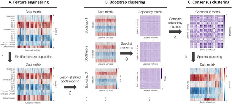
We studied 46 patients with histologically verified FCD Type II and 35 age- and sex-matched healthy controls. We applied consensus clustering, an unsupervised learning technique that identifies stable clusters based on bootstrap-aggregation, to 3 T multicontrast MRI (T1-weighted MRI and FLAIR) features of FCD normalized with respect to distributions in controls.

RESULTS

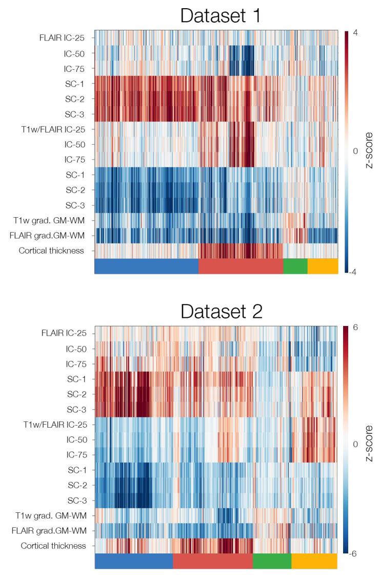
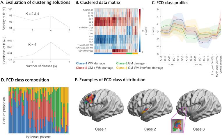
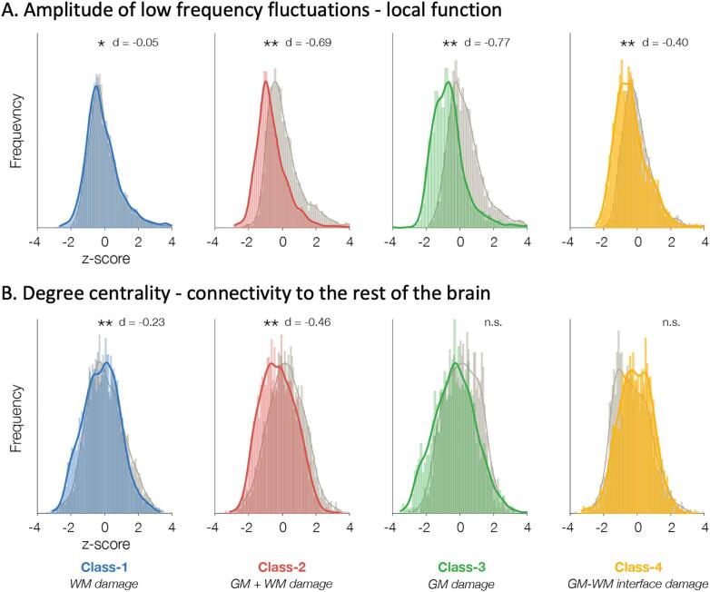
Lesions were parcellated into four classes with distinct structural profiles variably expressed within and across patients: Class-1 with isolated white matter (WM) damage; Class-2 combining grey matter (GM) and WM alterations; Class-3 with isolated GM damage; Class-4 with GM-WM interface anomalies. Class membership was replicated in two independent datasets. Classes with GM anomalies impacted local function (resting-state fMRI derived ALFF), while those with abnormal WM affected large-scale connectivity (assessed by degree centrality). Overall, MRI classes reflected typical histopathological FCD characteristics: Class-1 was associated with severe WM gliosis and interface blurring, Class-2 with severe GM dyslamination and moderate WM gliosis, Class-3 with moderate GM gliosis, Class-4 with mild interface blurring. A detection algorithm trained on class-informed data outperformed a class-naïve paradigm.

SIGNIFICANCE

Machine learning applied to widely available MRI contrasts uncovers FCD Type II variability at a mesoscopic scale and identifies tissue classes with distinct structural dimensions, functional and histopathological profiles. Integrating in vivo staging of FCD traits with automated lesion detection is likely to inform the development of novel personalized treatments.

摘要

目的

局灶性皮质发育不良(FCD)是最常见的致痫性发育畸形,也是手术可治疗癫痫的常见原因。虽然细胞和分子生物学数据表明 FCD 病变特征呈连续谱分布,但这一概念仍有待在体内验证。我们检验了这样一个假设,即应用于 MRI 的机器学习可在介观尺度上捕捉 FCD 病变的可变性。

方法

我们研究了 46 例经组织学证实的 FCD II 型患者和 35 名年龄和性别匹配的健康对照者。我们应用共识聚类(一种无监督学习技术,基于 bootstrap 聚合识别稳定的聚类),对 FCD 患者的 3T 多对比度 MRI(T1 加权 MRI 和 FLAIR)特征进行分析,这些特征经过与对照组分布归一化。

结果

病变被分割成四个具有不同结构特征的类别,这些特征在患者之间和内部表现出不同的表达:第 1 类为孤立的白质(WM)损伤;第 2 类为灰质(GM)和 WM 改变的组合;第 3 类为孤立的 GM 损伤;第 4 类为 GM-WM 界面异常。类别成员在两个独立数据集得到复制。具有 GM 异常的类别影响局部功能(静息态 fMRI 衍生的 ALFF),而具有异常 WM 的类别影响大尺度连通性(通过度中心性评估)。总的来说,MRI 类别反映了典型的组织病理学 FCD 特征:第 1 类与严重的 WM 胶质增生和界面模糊有关,第 2 类与严重的 GM 层状结构紊乱和中度 WM 胶质增生有关,第 3 类与中度 GM 胶质增生有关,第 4 类与轻度界面模糊有关。基于类别信息训练的检测算法优于类别未知的范式。

意义

应用于广泛可用的 MRI 对比的机器学习揭示了 FCD II 型在介观尺度上的可变性,并识别了具有不同结构维度、功能和组织病理学特征的组织类别。将 FCD 特征的体内分期与自动病变检测相结合,可能有助于开发新的个性化治疗方法。

https://cdn.ncbi.nlm.nih.gov/pmc/blobs/92fb/7520429/0aaa582c55b0/fx1.jpg
https://cdn.ncbi.nlm.nih.gov/pmc/blobs/92fb/7520429/5b6d38aa2037/gr1.jpg
https://cdn.ncbi.nlm.nih.gov/pmc/blobs/92fb/7520429/0a8e50e24c01/gr2.jpg
https://cdn.ncbi.nlm.nih.gov/pmc/blobs/92fb/7520429/21093951fa6b/gr3.jpg
https://cdn.ncbi.nlm.nih.gov/pmc/blobs/92fb/7520429/0aaa582c55b0/fx1.jpg
https://cdn.ncbi.nlm.nih.gov/pmc/blobs/92fb/7520429/5b6d38aa2037/gr1.jpg
https://cdn.ncbi.nlm.nih.gov/pmc/blobs/92fb/7520429/0a8e50e24c01/gr2.jpg
https://cdn.ncbi.nlm.nih.gov/pmc/blobs/92fb/7520429/21093951fa6b/gr3.jpg
https://cdn.ncbi.nlm.nih.gov/pmc/blobs/92fb/7520429/0aaa582c55b0/fx1.jpg

相似文献

1
Unsupervised machine learning reveals lesional variability in focal cortical dysplasia at mesoscopic scale.无监督机器学习揭示了微观尺度下局灶性皮质发育不良的病变变异性。
Neuroimage Clin. 2020;28:102438. doi: 10.1016/j.nicl.2020.102438. Epub 2020 Sep 18.
2
Gray-matter-specific MR imaging improves the detection of epileptogenic zones in focal cortical dysplasia: A new sequence called fluid and white matter suppression (FLAWS).灰质特异性磁共振成像提高局灶性皮质发育不良致痫灶的检出:一种新的序列,称为液体和白质抑制(FLAWS)。
Neuroimage Clin. 2018 Aug 11;20:388-397. doi: 10.1016/j.nicl.2018.08.010. eCollection 2018.
3
Multimodal MRI profiling of focal cortical dysplasia type II.II型局灶性皮质发育不良的多模态磁共振成像分析
Neurology. 2017 Feb 21;88(8):734-742. doi: 10.1212/WNL.0000000000003632. Epub 2017 Jan 27.
4
A connectome-based mechanistic model of focal cortical dysplasia.基于连接组学的局灶性皮质发育不良的机制模型。
Brain. 2019 Mar 1;142(3):688-699. doi: 10.1093/brain/awz009.
5
Automated detection of focal cortical dysplasia type II with surface-based magnetic resonance imaging postprocessing and machine learning.基于表面的磁共振成像后处理和机器学习自动检测 II 型局灶性皮质发育不良。
Epilepsia. 2018 May;59(5):982-992. doi: 10.1111/epi.14064. Epub 2018 Apr 10.
6
Combining magnetic resonance fingerprinting with voxel-based morphometric analysis to reduce false positives for focal cortical dysplasia detection.联合磁共振指纹技术与体素形态计量分析减少局灶性皮质发育不良检测的假阳性。
Epilepsia. 2024 Jun;65(6):1631-1643. doi: 10.1111/epi.17951. Epub 2024 Mar 21.
7
In vivo profiling of focal cortical dysplasia on high-resolution MRI with computational models.利用计算模型在高分辨率磁共振成像上对局灶性皮质发育异常进行体内分析。
Epilepsia. 2006 Jan;47(1):134-42. doi: 10.1111/j.1528-1167.2006.00379.x.
8
Cortical feature analysis and machine learning improves detection of "MRI-negative" focal cortical dysplasia.皮质特征分析和机器学习可提高对“MRI阴性”局灶性皮质发育异常的检测。
Epilepsy Behav. 2015 Jul;48:21-8. doi: 10.1016/j.yebeh.2015.04.055. Epub 2015 May 31.
9
Detection of cortical malformations using enhanced synthetic contrast images derived from quantitative T1 maps.利用基于定量 T1 图谱的增强合成对比图像检测皮质畸形。
NMR Biomed. 2020 Feb;33(2):e4203. doi: 10.1002/nbm.4203. Epub 2019 Dec 3.
10
Novel surface features for automated detection of focal cortical dysplasias in paediatric epilepsy.用于小儿癫痫中局灶性皮质发育异常自动检测的新型表面特征
Neuroimage Clin. 2016 Dec 30;14:18-27. doi: 10.1016/j.nicl.2016.12.030. eCollection 2017.

引用本文的文献

1
A nnU-Net-based automatic segmentation of FCD type II lesions in 3D FLAIR MRI images.基于nnU-Net的3D液体衰减反转恢复(FLAIR)磁共振成像(MRI)图像中II型局灶性皮质发育不良(FCD)病变的自动分割
Front Artif Intell. 2025 Jun 27;8:1601815. doi: 10.3389/frai.2025.1601815. eCollection 2025.
2
Matrix-assisted laser desorption/ionization mass spectrometry imaging as a new tool for molecular histopathology in epilepsy surgery.基质辅助激光解吸/电离质谱成像作为癫痫手术中分子组织病理学的一种新工具。
Epilepsia. 2024 Dec;65(12):3631-3643. doi: 10.1111/epi.18136. Epub 2024 Oct 5.
3
Mapping Functional Connectivity Signatures of Pharmacoresistant Focal Cortical Dysplasia-Related Epilepsy.

本文引用的文献

1
Somatic mosaicism and neurodevelopmental disease.体细胞镶嵌性与神经发育性疾病。
Nat Neurosci. 2018 Nov;21(11):1504-1514. doi: 10.1038/s41593-018-0257-3. Epub 2018 Oct 22.
2
Review: The international consensus classification of Focal Cortical Dysplasia - a critical update 2018.综述:《2018 年局灶性皮质发育不良国际共识分类——关键性更新》。
Neuropathol Appl Neurobiol. 2018 Feb;44(1):18-31. doi: 10.1111/nan.12462.
3
Review: Mechanistic target of rapamycin (mTOR) pathway, focal cortical dysplasia and epilepsy.综述:雷帕霉素靶蛋白(mTOR)通路、局灶性皮质发育不良和癫痫。
绘制药物难治性局灶性皮质发育不良相关癫痫的功能连接特征图谱。
Ann Neurol. 2024 Aug 27. doi: 10.1002/ana.27069.
4
Artificial intelligence in epilepsy - applications and pathways to the clinic.人工智能在癫痫中的应用及向临床应用的转化。
Nat Rev Neurol. 2024 Jun;20(6):319-336. doi: 10.1038/s41582-024-00965-9. Epub 2024 May 8.
5
Cortical surface analysis for focal cortical dysplasia diagnosis by using PET images.利用PET图像进行局灶性皮质发育不良诊断的皮质表面分析
Heliyon. 2023 Dec 12;10(1):e23605. doi: 10.1016/j.heliyon.2023.e23605. eCollection 2024 Jan 15.
6
Automatic Detection of Focal Cortical Dysplasia Using MRI: A Systematic Review.基于 MRI 的局灶性皮质发育不良自动检测:系统综述。
Sensors (Basel). 2023 Aug 10;23(16):7072. doi: 10.3390/s23167072.
7
Machine learning in neuroimaging: from research to clinical practice.神经影像学中的机器学习:从研究到临床实践。
Radiologie (Heidelb). 2022 Dec;62(Suppl 1):1-10. doi: 10.1007/s00117-022-01051-1. Epub 2022 Aug 31.
8
Interpretable surface-based detection of focal cortical dysplasias: a Multi-centre Epilepsy Lesion Detection study.基于表面的可解释性局灶性皮质发育不良检测:一项多中心癫痫病灶检测研究。
Brain. 2022 Nov 21;145(11):3859-3871. doi: 10.1093/brain/awac224.
9
Clinical Application of Machine Learning Models for Brain Imaging in Epilepsy: A Review.机器学习模型在癫痫脑成像中的临床应用:综述
Front Neurosci. 2021 Jun 22;15:684825. doi: 10.3389/fnins.2021.684825. eCollection 2021.
10
Recent Advances in Neuroimaging of Epilepsy.癫痫的神经影像学最新进展。
Neurotherapeutics. 2021 Apr;18(2):811-826. doi: 10.1007/s13311-021-01049-y. Epub 2021 May 3.
Neuropathol Appl Neurobiol. 2018 Feb;44(1):6-17. doi: 10.1111/nan.12463.
4
Multidimensional Neuroanatomical Subtyping of Autism Spectrum Disorder.自闭症谱系障碍的多维神经解剖学亚型。
Cereb Cortex. 2018 Oct 1;28(10):3578-3588. doi: 10.1093/cercor/bhx229.
5
Preferential susceptibility of limbic cortices to microstructural damage in temporal lobe epilepsy: A quantitative T1 mapping study.颞叶癫痫患者边缘皮质对结构损伤的易感性:一项定量 T1 映射研究。
Neuroimage. 2018 Nov 15;182:294-303. doi: 10.1016/j.neuroimage.2017.06.002. Epub 2017 Jun 3.
6
Characterization of focal cortical dysplasia with balloon cells by layer-specific markers: Evidence for differential vulnerability of interneurons.利用层特异性标志物对伴有气球样细胞的局灶性皮质发育异常进行特征分析:中间神经元不同易损性的证据
Epilepsia. 2017 Apr;58(4):635-645. doi: 10.1111/epi.13690. Epub 2017 Feb 16.
7
Focal Cortical Dysplasia: Gene Mutations, Cell Signaling, and Therapeutic Implications.局灶性皮质发育不良:基因突变、细胞信号传导及治疗意义。
Annu Rev Pathol. 2017 Jan 24;12:547-571. doi: 10.1146/annurev-pathol-052016-100138.
8
Resting-state connectivity biomarkers define neurophysiological subtypes of depression.静息态连接生物标志物定义了抑郁症的神经生理亚型。
Nat Med. 2017 Jan;23(1):28-38. doi: 10.1038/nm.4246. Epub 2016 Dec 5.
9
Impaired oligodendroglial turnover is associated with myelin pathology in focal cortical dysplasia and tuberous sclerosis complex.少突胶质细胞更新受损与局灶性皮质发育不良和结节性硬化症中的髓鞘病理相关。
Brain Pathol. 2017 Nov;27(6):770-780. doi: 10.1111/bpa.12452. Epub 2017 Feb 9.
10
Bayesian model reveals latent atrophy factors with dissociable cognitive trajectories in Alzheimer's disease.贝叶斯模型揭示了阿尔茨海默病中具有可分离认知轨迹的潜在萎缩因素。
Proc Natl Acad Sci U S A. 2016 Oct 18;113(42):E6535-E6544. doi: 10.1073/pnas.1611073113. Epub 2016 Oct 4.