Center for Biomedical Imaging and Neuromodulation, Nathan Kline Institute, Orangeburg, NY, USA.
Department of Biomedical Imaging and Image-guided Therapy, Computational Imaging Research Lab, Medical University of Vienna, Währinger Gürtel 18-20, 1090, Vienna, Austria.
Radiologie (Heidelb). 2022 Dec;62(Suppl 1):1-10. doi: 10.1007/s00117-022-01051-1. Epub 2022 Aug 31.
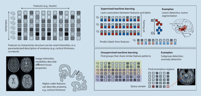
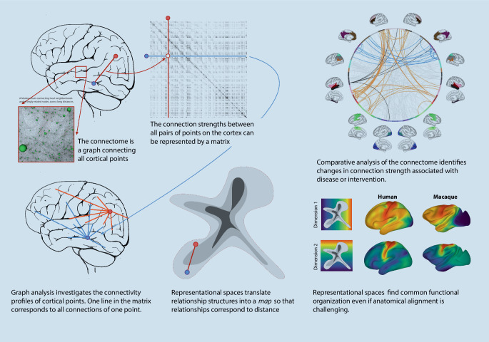
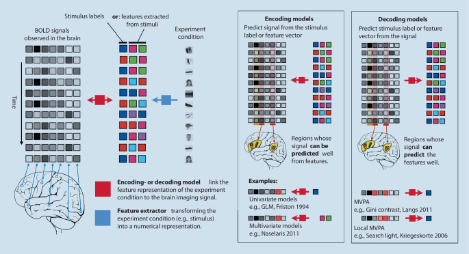
Neuroimaging is critical in clinical care and research, enabling us to investigate the brain in health and disease. There is a complex link between the brain's morphological structure, physiological architecture, and the corresponding imaging characteristics. The shape, function, and relationships between various brain areas change during development and throughout life, disease, and recovery. Like few other areas, neuroimaging benefits from advanced analysis techniques to fully exploit imaging data for studying the brain and its function. Recently, machine learning has started to contribute (a) to anatomical measurements, detection, segmentation, and quantification of lesions and disease patterns, (b) to the rapid identification of acute conditions such as stroke, or (c) to the tracking of imaging changes over time. As our ability to image and analyze the brain advances, so does our understanding of its intricate relationships and their role in therapeutic decision-making. Here, we review the current state of the art in using machine learning techniques to exploit neuroimaging data for clinical care and research, providing an overview of clinical applications and their contribution to fundamental computational neuroscience.
神经影像学在临床护理和研究中至关重要,使我们能够研究健康和疾病中的大脑。大脑的形态结构、生理结构和相应的成像特征之间存在着复杂的联系。在发育过程中以及在整个生命周期中,疾病和康复期间,大脑的各种区域的形状、功能和关系都在发生变化。像其他很少的领域一样,神经影像学受益于先进的分析技术,以充分利用成像数据来研究大脑及其功能。最近,机器学习开始为以下方面做出贡献:(a) 对解剖学测量、病变和疾病模式的检测、分割和量化,(b) 对中风等急性疾病的快速识别,或 (c) 对随时间推移的成像变化的跟踪。随着我们对大脑成像和分析能力的提高,我们对其复杂关系及其在治疗决策中的作用的理解也在不断提高。在这里,我们回顾了使用机器学习技术来利用神经影像学数据进行临床护理和研究的最新进展,概述了临床应用及其对基础计算神经科学的贡献。