Cagnoli Cinzia, De Santis Dalia, Caccia Claudio, Bongarzone Italia, Capitoli Giulia, Rossini Laura, Rizzi Michele, Deleo Francesco, Tassi Laura, de Curtis Marco, Garbelli Rita
Epilepsy Unit, Fondazione IRCCS Istituto Neurologico Carlo Besta, Milan, Italy.
Medical Genetics and Neurogenetic Unit, Fondazione IRCCS Istituto Neurologico Carlo Besta, Milan, Italy.
Epilepsia. 2024 Dec;65(12):3631-3643. doi: 10.1111/epi.18136. Epub 2024 Oct 5.
Epilepsy surgery is a treatment option for patients with seizures that do not respond to pharmacotherapy. The histopathological characterization of the resected tissue has an important prognostic value to define postoperative seizure outcome in these patients. However, the diagnostic classification process based on microscopic assessment remains challenging, particularly in the case of focal cortical dysplasia (FCD). Imaging mass spectrometry is a spatial omics technique that could improve tissue phenotyping and patient stratification by investigating hundreds of biomolecules within a single tissue sample, without the need for target-specific reagents.
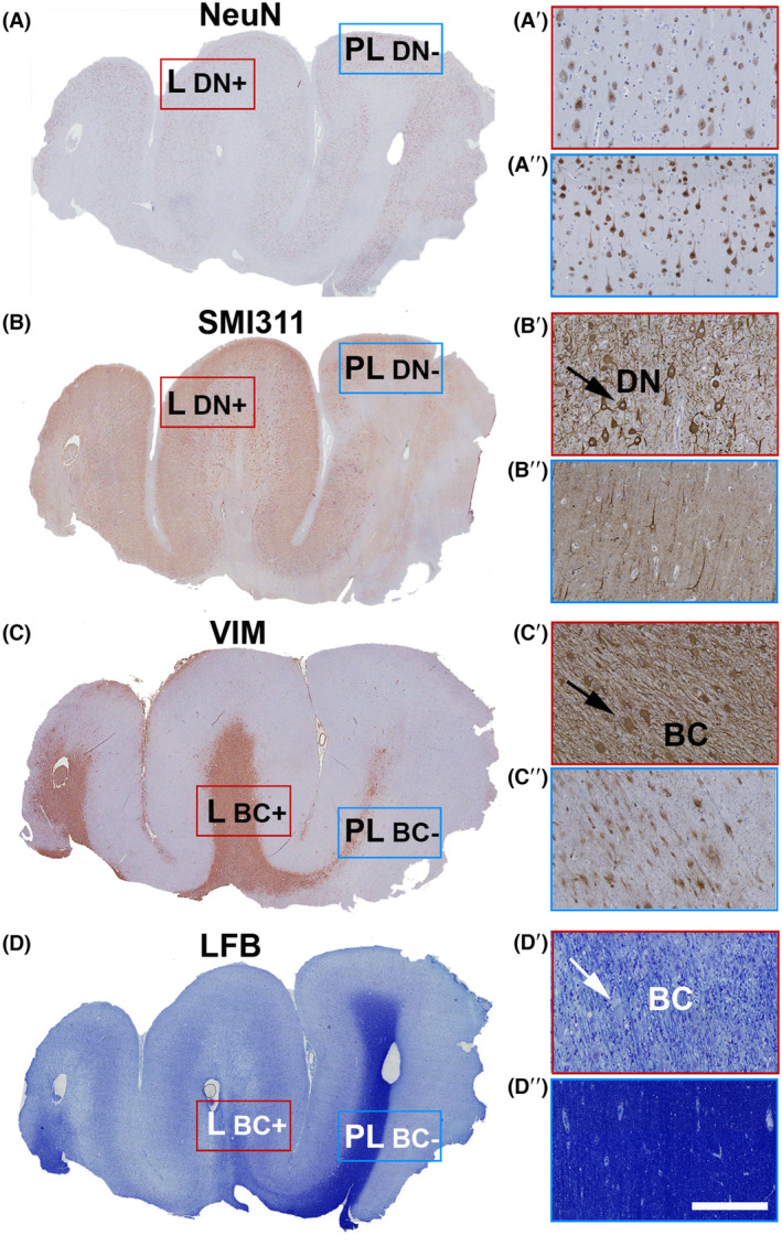
An in situ proteomic technique called matrix-assisted laser desorption/ionization mass spectrometry imaging (MALDI-MSI) is here investigated as a potential new tool to expand conventional diagnosis on standard paraffin brain tissue sections. Unsupervised and region of interest-based MALDI-MSI analyses of sections from 10 FCD type IIb (FCDIIb) cases were performed, and the results were validated by immunohistochemistry.
MALDI-MSI identified distinct histopathological features and the boundaries of the dysplastic lesion. The capability to visualize the spatial distribution of well-known diagnostic markers enabling multiplex measurements on single tissue sections was demonstrated. Finally, a fingerprint list of potential discriminant peptides that distinguish FCD core from peri-FCD tissue was generated.
This is the first study that explores the potential application of MALDI-MSI in epilepsy postsurgery fixed tissue, by utilizing the well-characterized FCDIIb features as a model. Extending these preliminary analyses to a larger cohort of patients will generate spectral libraries of molecular signatures that discriminate tissue features and will contribute to patient phenotyping.
癫痫手术是药物治疗无效的癫痫患者的一种治疗选择。切除组织的组织病理学特征对于确定这些患者术后癫痫发作结果具有重要的预后价值。然而,基于显微镜评估的诊断分类过程仍然具有挑战性,尤其是在局灶性皮质发育不良(FCD)的情况下。成像质谱是一种空间组学技术,它可以通过研究单个组织样本中的数百种生物分子来改善组织表型分析和患者分层,而无需针对特定靶点的试剂。
本文研究了一种称为基质辅助激光解吸/电离质谱成像(MALDI-MSI)的原位蛋白质组学技术,作为一种潜在的新工具,用于扩展对标准石蜡脑组织切片的传统诊断。对10例IIb型局灶性皮质发育不良(FCDIIb)病例的切片进行了无监督和基于感兴趣区域的MALDI-MSI分析,并通过免疫组织化学对结果进行了验证。
MALDI-MSI识别出了不同的组织病理学特征和发育异常病变的边界。展示了可视化已知诊断标志物空间分布的能力,从而能够在单个组织切片上进行多重测量。最后,生成了一份区分FCD核心与FCD周围组织的潜在判别肽指纹列表。
这是第一项以特征明确的FCDIIb特征为模型,探索MALDI-MSI在癫痫手术后固定组织中的潜在应用的研究。将这些初步分析扩展到更大的患者队列将生成区分组织特征的分子特征光谱库,并有助于患者表型分析。