Department of Nutritional Science, Faculty of Applied Bioscience, Tokyo University of Agriculture, 1-1-1 Sakuragaoka, Setagaya-ku, Tokyo 156-8502, Japan.
Department of Cell Biological Science, Faculty of Advanced Life Science, Hokkaido University, Kita-21, Nishi-11, Kita-ku, Sapporo, Hokkaido 001-0021, Japan.
Toxins (Basel). 2020 Sep 24;12(10):610. doi: 10.3390/toxins12100610.
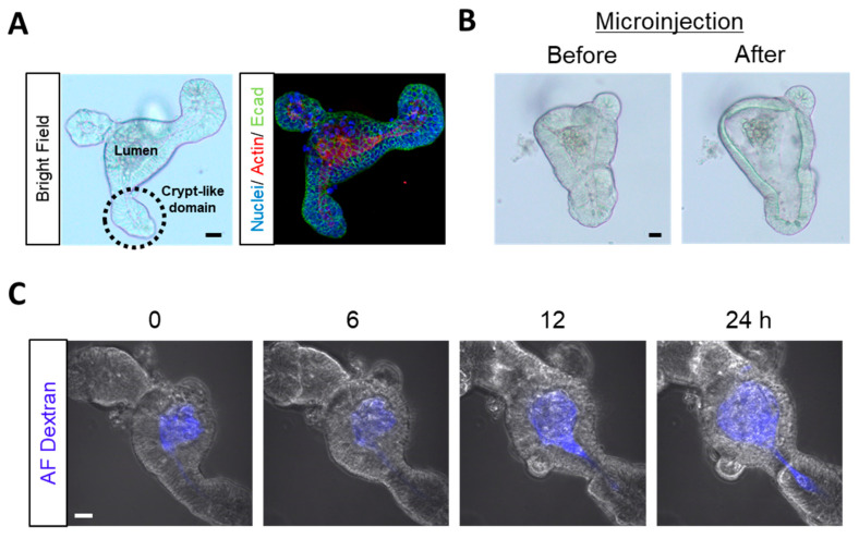
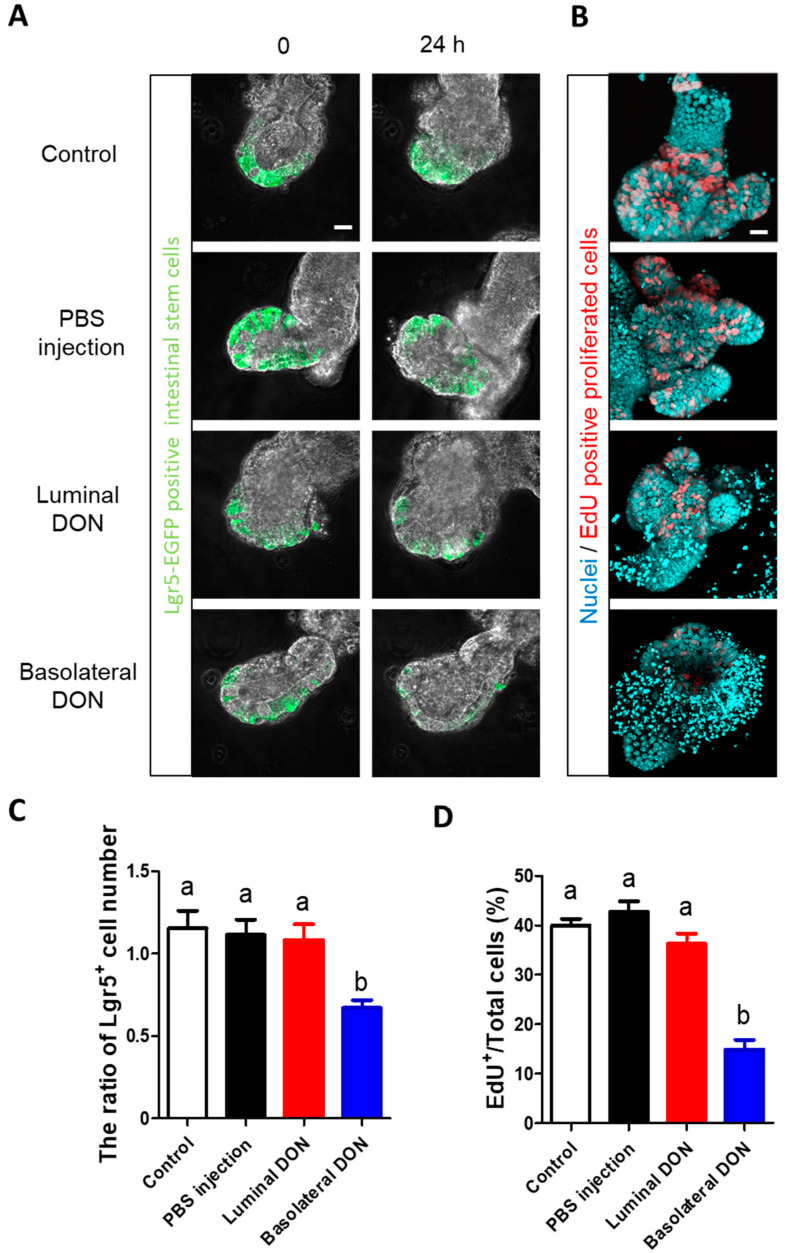
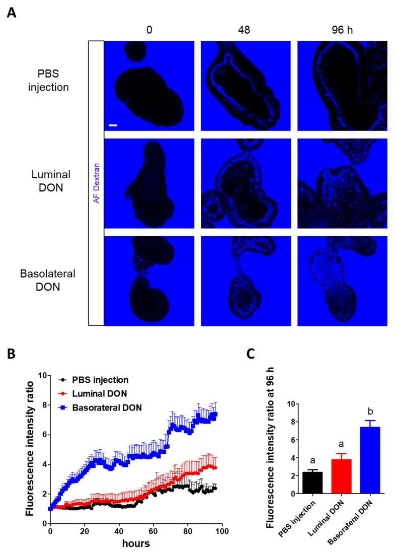
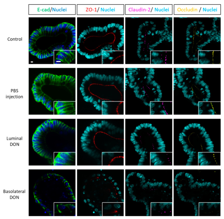
The different effects of deoxynivalenol (DON) on intestinal barrier and stem cells by its route of exposure remain less known. We explored the toxic effects of DON on intestinal barrier functions and stem cells after DON microinjection (luminal exposure) or addition to a culture medium (basolateral exposure) using three-dimensional mouse intestinal organoids (enteroids). The influx test using fluorescein-labeled dextran showed that basolateral DON exposure (1 micromolar (µM) disrupted intestinal barrier functions in enteroids compared with luminal DON exposure at the same concentration. Moreover, an immunofluorescence experiment of intestinal epithelial proteins, such as E-cadherin, claudin, zonula occludens-1 (ZO-1), and occludin, exhibited that only basolateral DON exposure broke down intestinal epithelial integrity. A time-lapse analysis using enteroids from leucine-rich repeat-containing G-protein-coupled receptor 5 (Lgr5)-enhanced green fluorescence protein (EGFP) transgenic mice and 5-ethynyl-2-deoxyuridine (EdU) assay indicated that only the basolateral DON exposure, but not luminal DON exposure, suppressed Lgr5 stem cell count and proliferative cell ratio, respectively. These results revealed that basolateral DON exposure has larger impacts on intestinal barrier function and stem cells than luminal DON exposure. This is the first report that DON had different impacts on intestinal stem cells depending on the administration route. In addition, RNA sequencing analysis showed different expression of genes among enteroids after basolateral and luminal DON exposure.
脱氧雪腐镰刀菌烯醇(DON)通过不同途径暴露对肠道屏障和干细胞的影响尚不清楚。我们使用三维小鼠肠类器官(肠类器官)探索了 DON 经微注射(腔暴露)或添加至培养基(基底外侧暴露)后对肠道屏障功能和干细胞的毒性作用。使用荧光素标记的葡聚糖的流入试验表明,与相同浓度的腔暴露相比,基底外侧 DON 暴露(1 微摩尔(µM)破坏了肠类器官的肠道屏障功能。此外,肠上皮蛋白(如 E-钙黏蛋白、闭合蛋白、紧密连接蛋白-1(ZO-1)和闭合蛋白)的免疫荧光实验表明,只有基底外侧 DON 暴露破坏了肠上皮完整性。使用来自富含亮氨酸重复序列的 G 蛋白偶联受体 5(Lgr5)-增强型绿色荧光蛋白(EGFP)转基因小鼠的肠类器官和 5-乙炔基-2-脱氧尿苷(EdU)测定的延时分析表明,只有基底外侧 DON 暴露分别抑制了 Lgr5 干细胞计数和增殖细胞比例,而腔 DON 暴露则没有。这些结果表明,基底外侧 DON 暴露对肠道屏障功能和干细胞的影响大于腔 DON 暴露。这是首次报道 DON 对肠道干细胞的影响取决于给药途径。此外,RNA 测序分析表明,基底外侧和腔 DON 暴露后肠类器官中基因的表达不同。