• 文献检索
  • 文档翻译
  • 深度研究
  • 学术资讯
  • Suppr Zotero 插件Zotero 插件
  • 邀请有礼
  • 套餐&价格
  • 历史记录
应用&插件
Suppr Zotero 插件Zotero 插件浏览器插件Mac 客户端Windows 客户端微信小程序
定价
高级版会员购买积分包购买API积分包
服务
文献检索文档翻译深度研究API 文档MCP 服务
关于我们
关于 Suppr公司介绍联系我们用户协议隐私条款
关注我们

Suppr 超能文献

核心技术专利:CN118964589B侵权必究
粤ICP备2023148730 号-1Suppr @ 2026

文献检索

告别复杂PubMed语法,用中文像聊天一样搜索,搜遍4000万医学文献。AI智能推荐,让科研检索更轻松。

立即免费搜索

文件翻译

保留排版,准确专业,支持PDF/Word/PPT等文件格式,支持 12+语言互译。

免费翻译文档

深度研究

AI帮你快速写综述,25分钟生成高质量综述,智能提取关键信息,辅助科研写作。

立即免费体验

MAIT 细胞的激活与 COVID-19 疾病严重程度相关。

MAIT cell activation and dynamics associated with COVID-19 disease severity.

机构信息

Center for Infectious Medicine, Department of Medicine Huddinge, Karolinska Institutet, Karolinska University Hospital, Stockholm, Sweden.

Department of Infectious Diseases, Karolinska University Hospital, Stockholm, Sweden.

出版信息

Sci Immunol. 2020 Sep 28;5(51). doi: 10.1126/sciimmunol.abe1670.

DOI:10.1126/sciimmunol.abe1670
PMID:32989174
原文链接:https://pmc.ncbi.nlm.nih.gov/articles/PMC7857393/
Abstract

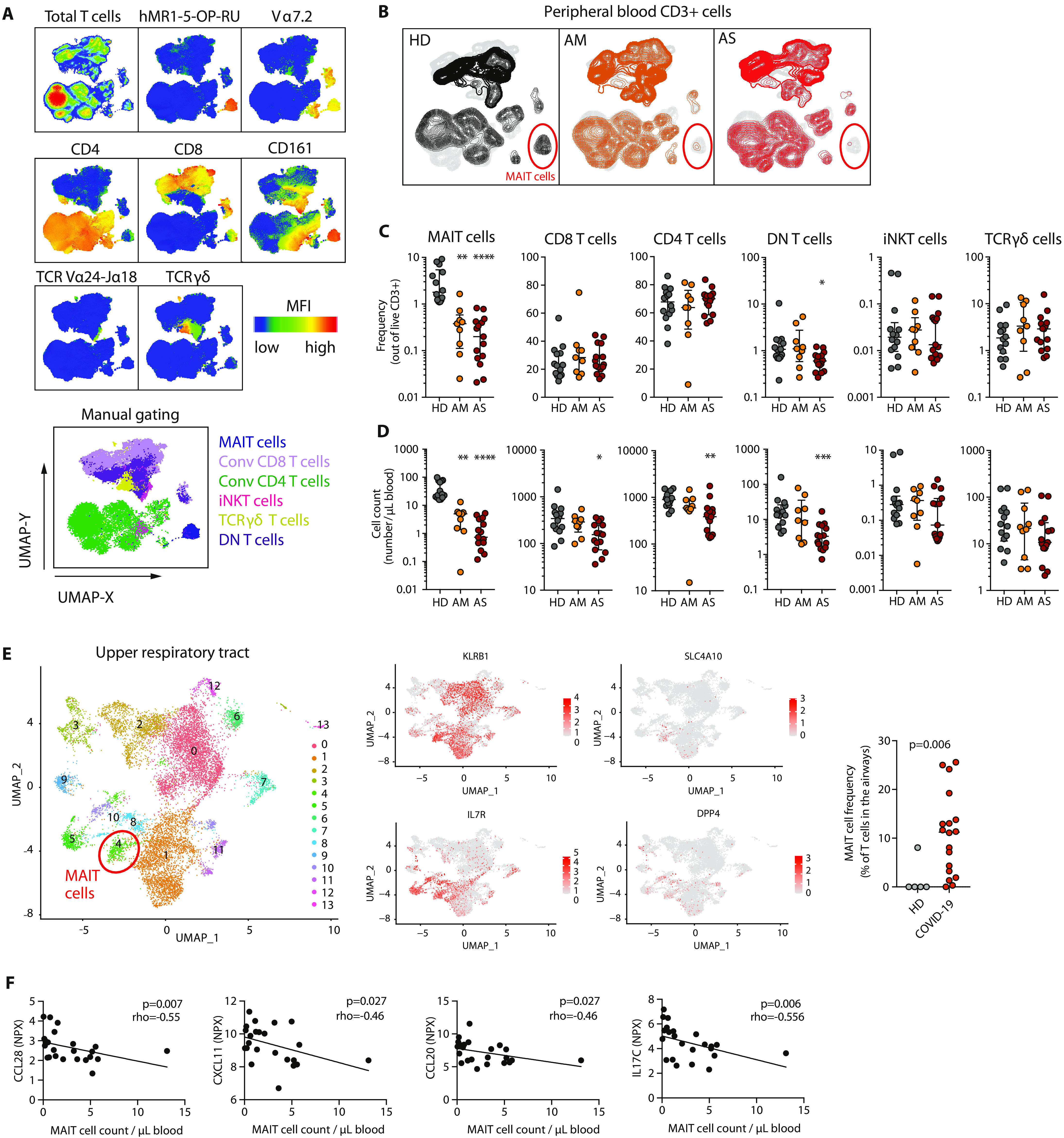
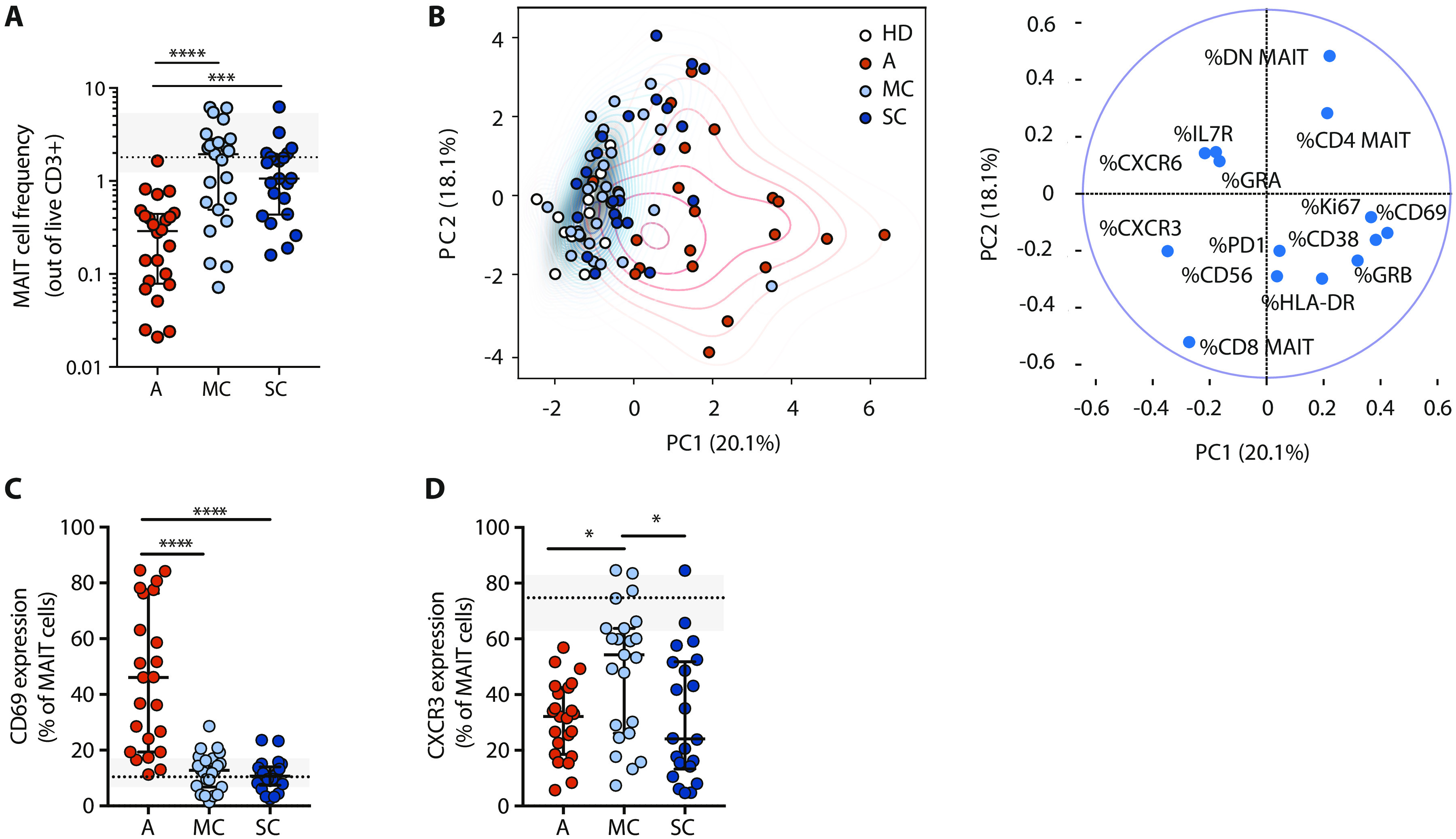
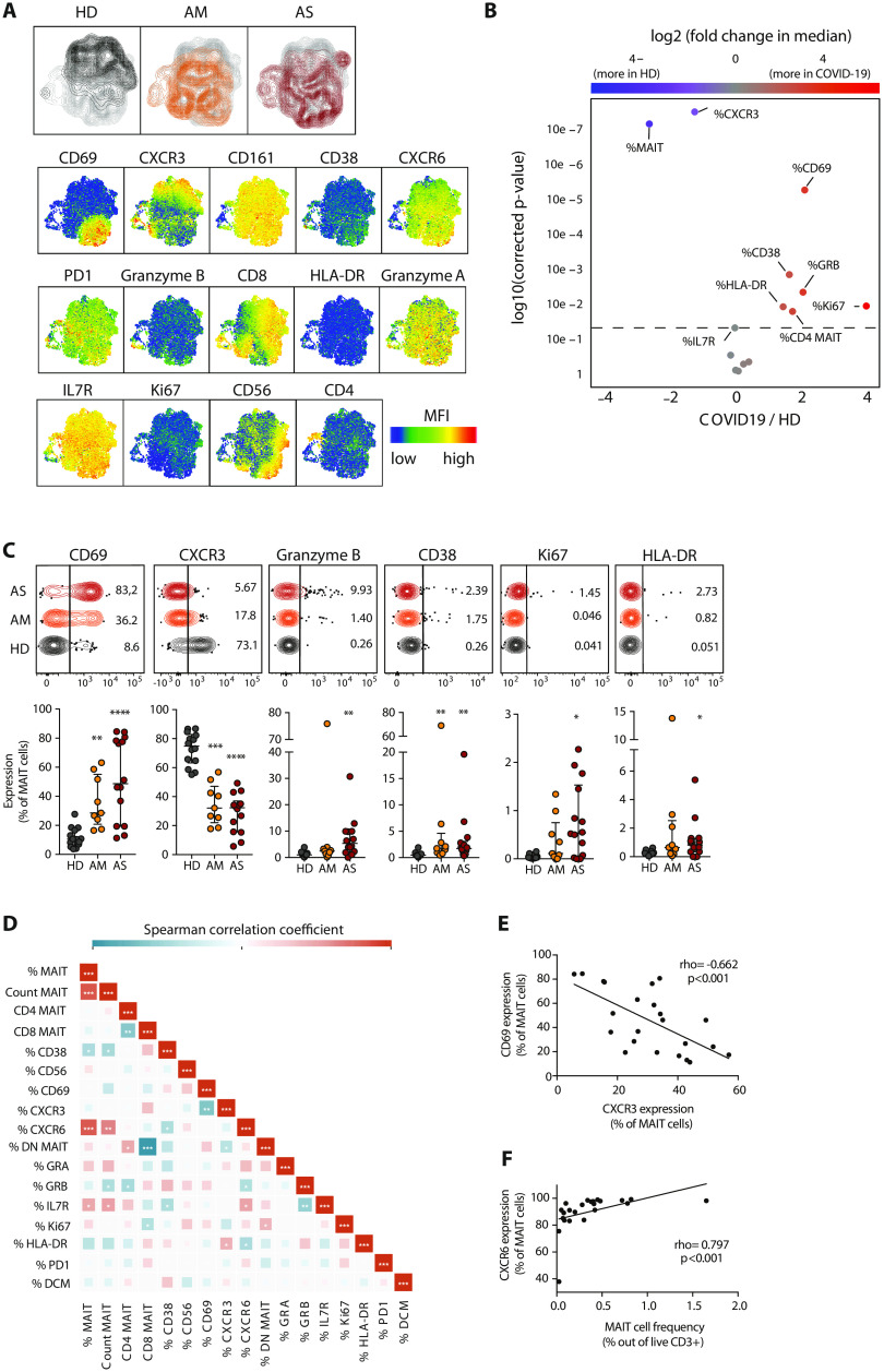
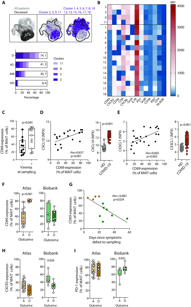
Severe COVID-19 is characterized by excessive inflammation of the lower airways. The balance of protective versus pathological immune responses in COVID-19 is incompletely understood. Mucosa-associated invariant T (MAIT) cells are antimicrobial T cells that recognize bacterial metabolites, and can also function as innate-like sensors and mediators of antiviral responses. Here, we investigated the MAIT cell compartment in COVID-19 patients with moderate and severe disease, as well as in convalescence. We show profound and preferential decline in MAIT cells in the circulation of patients with active disease paired with strong activation. Furthermore, transcriptomic analyses indicated significant MAIT cell enrichment and pro-inflammatory IL-17A bias in the airways. Unsupervised analysis identified MAIT cell CD69 and CXCR3 immunotypes associated with poor clinical outcome. MAIT cell levels normalized in the convalescent phase, consistent with dynamic recruitment to the tissues and later release back into the circulation when disease is resolved. These findings indicate that MAIT cells are engaged in the immune response against SARS-CoV-2 and suggest their possible involvement in COVID-19 immunopathogenesis.

摘要

严重的 COVID-19 的特征是下呼吸道过度炎症。COVID-19 中保护性免疫反应与病理性免疫反应之间的平衡尚未完全清楚。黏膜相关不变 T(MAIT)细胞是识别细菌代谢物的抗菌 T 细胞,也可以作为先天样传感器和抗病毒反应的介质发挥作用。在这里,我们研究了中度和重度疾病以及恢复期 COVID-19 患者的 MAIT 细胞区室。我们发现,与强烈激活配对的活动性疾病患者的循环中 MAIT 细胞明显减少并呈优先减少趋势。此外,转录组分析表明,气道中 MAIT 细胞的富集和促炎的 IL-17A 偏向明显。无监督分析确定了与不良临床结局相关的 MAIT 细胞 CD69 和 CXCR3 免疫表型。在恢复期,MAIT 细胞水平恢复正常,这与疾病消退时 MAIT 细胞向组织中的动态募集以及随后释放回循环一致。这些发现表明 MAIT 细胞参与了针对 SARS-CoV-2 的免疫反应,并提示它们可能参与了 COVID-19 的免疫发病机制。

https://cdn.ncbi.nlm.nih.gov/pmc/blobs/3641/7857393/becebd06ca31/abe1670-F4.jpg
https://cdn.ncbi.nlm.nih.gov/pmc/blobs/3641/7857393/56f70d66b881/abe1670-F1.jpg
https://cdn.ncbi.nlm.nih.gov/pmc/blobs/3641/7857393/b970c16a0693/abe1670-F2.jpg
https://cdn.ncbi.nlm.nih.gov/pmc/blobs/3641/7857393/64b534850687/abe1670-F3.jpg
https://cdn.ncbi.nlm.nih.gov/pmc/blobs/3641/7857393/becebd06ca31/abe1670-F4.jpg

相似文献

1
MAIT cell activation and dynamics associated with COVID-19 disease severity.MAIT 细胞的激活与 COVID-19 疾病严重程度相关。
Sci Immunol. 2020 Sep 28;5(51). doi: 10.1126/sciimmunol.abe1670.
2
MAIT Cells in COVID-19: Heroes, Villains, or Both?新冠病毒肺炎中的黏膜相关恒定T细胞:英雄、反派,还是兼而有之?
Crit Rev Immunol. 2020;40(2):173-184. doi: 10.1615/CritRevImmunol.2020034943.
3
Phenotypical and functional alteration of unconventional T cells in severe COVID-19 patients.在重症 COVID-19 患者中非常规 T 细胞的表型和功能改变。
J Exp Med. 2020 Dec 7;217(12). doi: 10.1084/jem.20200872.
4
Mucosal-Associated Invariant T (MAIT) Cells Are Highly Activated and Functionally Impaired in COVID-19 Patients.黏膜相关不变 T(MAIT)细胞在 COVID-19 患者中高度激活且功能受损。
Viruses. 2021 Feb 3;13(2):241. doi: 10.3390/v13020241.
5
MAIT cells are activated and accumulated in the inflamed mucosa of ulcerative colitis.黏膜相关恒定T细胞(MAIT细胞)在溃疡性结肠炎的炎症黏膜中被激活并积聚。
J Gastroenterol Hepatol. 2016 May;31(5):965-72. doi: 10.1111/jgh.13242.
6
Activation and Impaired Tumor Necrosis Factor-α Production of Circulating Mucosal-Associated Invariant T Cells in Patients with Trauma.创伤患者循环黏膜相关不变 T 细胞的激活和肿瘤坏死因子-α产生受损。
J Innate Immun. 2019;11(6):506-515. doi: 10.1159/000499343. Epub 2019 May 14.
7
Single-cell landscape of immunological responses in patients with COVID-19.COVID-19 患者免疫反应的单细胞景观。
Nat Immunol. 2020 Sep;21(9):1107-1118. doi: 10.1038/s41590-020-0762-x. Epub 2020 Aug 12.
8
Tissue-resident mucosal-associated invariant T (MAIT) cells in the human kidney represent a functionally distinct subset.人类肾脏组织驻留的黏膜相关恒定 T(MAIT)细胞代表了一个功能独特的亚群。
Eur J Immunol. 2020 Nov;50(11):1783-1797. doi: 10.1002/eji.202048644. Epub 2020 Aug 6.
9
Human mucosal-associated invariant T cells contribute to antiviral influenza immunity via IL-18-dependent activation.人类黏膜相关恒定T细胞通过依赖白细胞介素-18的激活作用来促进抗流感病毒免疫。
Proc Natl Acad Sci U S A. 2016 Sep 6;113(36):10133-8. doi: 10.1073/pnas.1610750113. Epub 2016 Aug 19.
10
Mucosal-Associated Invariant T Cells Develop an Innate-Like Transcriptomic Program in Anti-mycobacterial Responses.黏膜相关不变 T 细胞在抗分枝杆菌反应中形成固有样转录组程序。
Front Immunol. 2020 Jun 9;11:1136. doi: 10.3389/fimmu.2020.01136. eCollection 2020.

引用本文的文献

1
Persistent Immune Dysregulation during Post-Acute Sequelae of COVID-19 is Manifested in Antibodies Targeting Envelope and Nucleocapsid Proteins.新冠后遗症期间持续的免疫失调表现为针对包膜蛋白和核衣壳蛋白的抗体。
bioRxiv. 2025 Aug 19:2025.08.18.670908. doi: 10.1101/2025.08.18.670908.
2
High-dimensional single-cell phenotyping unveils persistent differences in immune cell profiles between severe and moderate seasonal influenza.高维单细胞表型分析揭示了重度和中度季节性流感之间免疫细胞谱的持续差异。
Front Immunol. 2025 Jul 22;16:1576861. doi: 10.3389/fimmu.2025.1576861. eCollection 2025.
3
Poxvirus Vectors Activate Human NK and MAIT Cells in a Type I Interferon, IL-18, and Monocyte-Dependent Manner.

本文引用的文献

1
Robust T Cell Immunity in Convalescent Individuals with Asymptomatic or Mild COVID-19.无症状或轻症 COVID-19 康复者体内具有强大的 T 细胞免疫。
Cell. 2020 Oct 1;183(1):158-168.e14. doi: 10.1016/j.cell.2020.08.017. Epub 2020 Aug 14.
2
Phenotypical and functional alteration of unconventional T cells in severe COVID-19 patients.在重症 COVID-19 患者中非常规 T 细胞的表型和功能改变。
J Exp Med. 2020 Dec 7;217(12). doi: 10.1084/jem.20200872.
3
Natural killer cell immunotypes related to COVID-19 disease severity.与 COVID-19 疾病严重程度相关的自然杀伤细胞免疫表型。
痘病毒载体以I型干扰素、白细胞介素-18和单核细胞依赖的方式激活人自然杀伤细胞和黏膜相关恒定T细胞。
J Immunol Res. 2025 Jul 12;2025:1203141. doi: 10.1155/jimr/1203141. eCollection 2025.
4
Single-cell RNA-sequencing highlights a curtailed NK cell function in convalescent COVID-19 pregnant women.单细胞RNA测序揭示了新冠康复期孕妇自然杀伤细胞功能的减弱。
Front Immunol. 2025 Jun 30;16:1560391. doi: 10.3389/fimmu.2025.1560391. eCollection 2025.
5
Temporal multi-omics analysis of COVID-19 in end-stage kidney disease.终末期肾病患者新冠病毒病的时间多组学分析
Cell Genom. 2025 Aug 13;5(8):100918. doi: 10.1016/j.xgen.2025.100918. Epub 2025 Jun 17.
6
Control of MAIT cell functions by cytokines in health and disease.健康与疾病状态下细胞因子对黏膜相关恒定T细胞(MAIT细胞)功能的调控
Front Immunol. 2025 May 30;16:1594712. doi: 10.3389/fimmu.2025.1594712. eCollection 2025.
7
Immune responses to SARS-CoV-2 infection in Humans and ACE2 humanized mice.人类和ACE2人源化小鼠对SARS-CoV-2感染的免疫反应。
Fundam Res. 2021 Mar;1(2):124-130. doi: 10.1016/j.fmre.2021.03.001. Epub 2021 Mar 3.
8
Unveiling the role of the upper respiratory tract microbiome in susceptibility and severity to COVID-19.揭示上呼吸道微生物群在新冠病毒易感性和严重程度中的作用。
Front Cell Infect Microbiol. 2025 May 13;15:1531084. doi: 10.3389/fcimb.2025.1531084. eCollection 2025.
9
Cellular and Molecular Atlas of Peripheral Blood Mononuclear Cells from a Pregnant Woman After Recovery from COVID-19.新冠康复后孕妇外周血单个核细胞的细胞和分子图谱
Matern Fetal Med. 2023 Apr 24;5(2):88-96. doi: 10.1097/FM9.0000000000000190. eCollection 2023 Apr.
10
The 15-Year Survival Advantage: Immune Resilience as a Salutogenic Force in Healthy Aging.15年生存优势:免疫弹性作为健康衰老中的一种健康促进力量。
Aging Cell. 2025 Apr 23:e70063. doi: 10.1111/acel.70063.
Sci Immunol. 2020 Aug 21;5(50). doi: 10.1126/sciimmunol.abd6832.
4
Absence of mucosal-associated invariant T cells in a person with a homozygous point mutation in .在一个纯合点突变的个体中缺失黏膜相关不变 T 细胞。
Sci Immunol. 2020 Jul 24;5(49). doi: 10.1126/sciimmunol.abc9492.
5
Deep immune profiling of COVID-19 patients reveals distinct immunotypes with therapeutic implications.深度免疫剖析 COVID-19 患者,揭示具有治疗意义的不同免疫类型。
Science. 2020 Sep 4;369(6508). doi: 10.1126/science.abc8511. Epub 2020 Jul 15.
6
Comprehensive mapping of immune perturbations associated with severe COVID-19.全面绘制与严重 COVID-19 相关的免疫扰动图谱。
Sci Immunol. 2020 Jul 15;5(49). doi: 10.1126/sciimmunol.abd7114.
7
SARS-CoV-2-specific T cell immunity in cases of COVID-19 and SARS, and uninfected controls.COVID-19 和 SARS 病例以及未感染对照者的 SARS-CoV-2 特异性 T 细胞免疫。
Nature. 2020 Aug;584(7821):457-462. doi: 10.1038/s41586-020-2550-z. Epub 2020 Jul 15.
8
Immunophenotyping of COVID-19 and influenza highlights the role of type I interferons in development of severe COVID-19.COVID-19 和流感的免疫表型分析突出了 I 型干扰素在 COVID-19 重症发展中的作用。
Sci Immunol. 2020 Jul 10;5(49). doi: 10.1126/sciimmunol.abd1554.
9
Marked T cell activation, senescence, exhaustion and skewing towards TH17 in patients with COVID-19 pneumonia.COVID-19 肺炎患者中标记的 T 细胞激活、衰老、衰竭和向 TH17 的倾斜。
Nat Commun. 2020 Jul 6;11(1):3434. doi: 10.1038/s41467-020-17292-4.
10
COVID-19 severity correlates with airway epithelium-immune cell interactions identified by single-cell analysis.单细胞分析鉴定的 COVID-19 严重程度与气道上皮-免疫细胞相互作用相关。
Nat Biotechnol. 2020 Aug;38(8):970-979. doi: 10.1038/s41587-020-0602-4. Epub 2020 Jun 26.