Division of Gerontology and Geriatric Medicine, Department of Medicine, University of Washington, Seattle, WA, USA.
Geriatric Research Education and Clinical Center, VA Puget Sound Health Care System, Seattle, WA, USA.
Fluids Barriers CNS. 2020 Sep 29;17(1):60. doi: 10.1186/s12987-020-00219-y.
The microvasculature (MV) of brains with Alzheimer's disease neuropathologic change (ADNC) and cerebral amyloid angiopathy (CAA), in the absence of concurrent pathologies (e.g., infarctions, Lewy bodies), is incompletely understood.
To analyze microvascular density, diameter and extracellular matrix (ECM) content in association with ADNC and CAA.
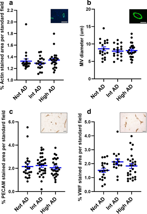
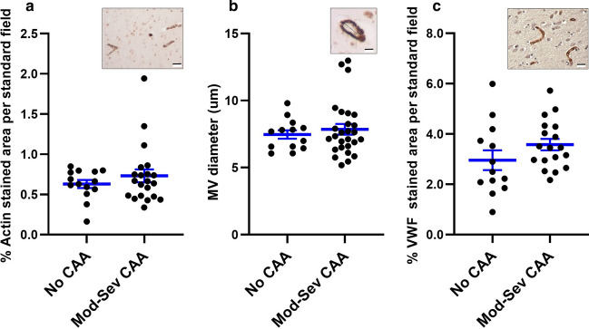
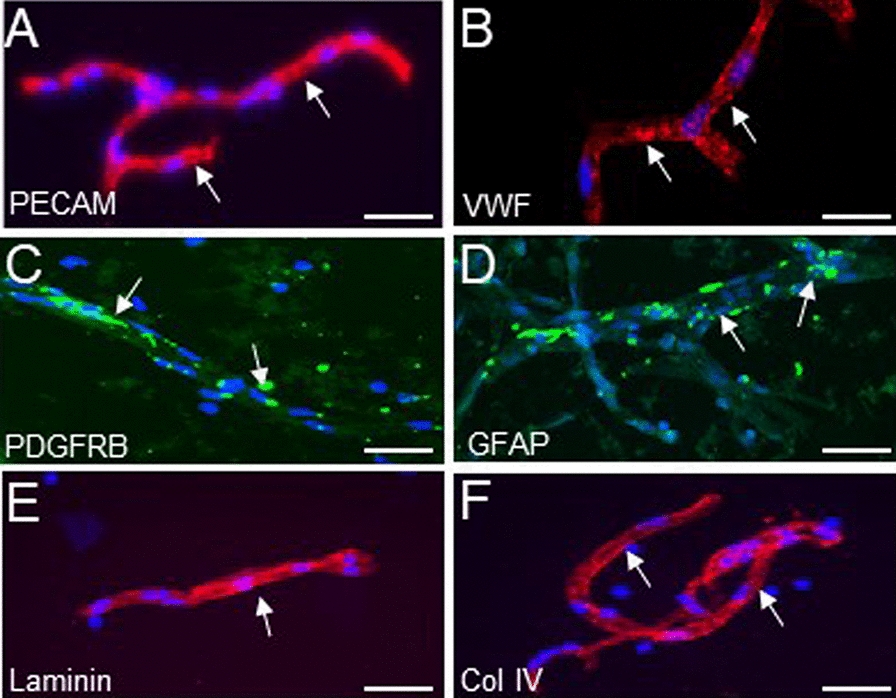
We examined samples of cerebral cortex and isolated brain microvasculature (MV) from subjects with the National Institute on Aging-Alzheimer's Association (NIA-AA) designations of not-, intermediate-, or high ADNC and from subjects with no CAA and moderate-severe CAA. Cases for all groups were selected with no major (territorial) strokes, ≤ 1 microinfarct in screening sections, and no Lewy body pathology. MV density and diameter were measured from cortical brain sections. Levels of basement membrane (BM) ECM components, the protein product of TNF-stimulated gene-6 (TSG-6), and the ubiquitous glycosaminoglycan hyaluronan (HA) were assayed by western blots or HA ELISA of MV lysates.
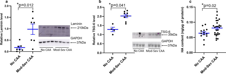
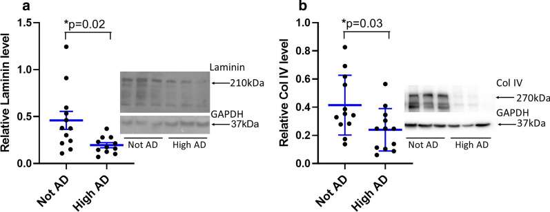
We found no significant changes in MV density or diameter among any of the groups. Levels of BM laminin and collagen IV (col IV) were lower in MV isolated from the high ADNC vs. not-ADNC groups. In contrast, BM laminin was significantly higher in MV from the moderate-severe CAA vs. the no CAA groups. TSG-6 and HA content were higher in the presence of both high ADNC and CAA, whereas levels of BM fibronectin and perlecan were similar among all groups.
Cortical MV density and diameter are not appreciably altered by ADNC or CAA. TSG-6 and HA are increased in both ADNC and CAA, with laminin and col IV decreased in the BM of high ADNC, but laminin increased in moderate-severe CAA. These results show that changes in the ECM occur in AD and CAA, but independently of one another, and likely reflect on the regional functioning of the brain microvasculature.
在没有合并病变(如梗死、路易体)的情况下,伴有阿尔茨海默病神经病理改变(ADNC)和脑淀粉样血管病(CAA)的大脑微血管(MV)尚未完全阐明。
分析与 ADNC 和 CAA 相关的微血管密度、直径和细胞外基质(ECM)含量。
我们检查了具有美国国家老龄化研究所-阿尔茨海默病协会(NIA-AA)指定的不伴有、中等程度伴有或高度伴有 ADNC 以及不伴有 CAA 和中重度 CAA 的受试者的大脑皮质和分离的脑微血管(MV)样本。所有组的病例均选择无主要(区域性)中风、筛查切片中≤1 个微梗死且无路易体病理学。从皮质脑切片测量 MV 密度和直径。通过 Western blot 或 MV 裂解物的透明质酸 ELISA 测定基底膜(BM)ECM 成分、肿瘤坏死因子刺激基因-6(TSG-6)的蛋白产物和无处不在的糖胺聚糖透明质酸(HA)的水平。
我们发现任何组之间 MV 密度或直径均无明显变化。与不伴有 ADNC 的组相比,高 ADNC 组的 MV 中 BM 层粘连蛋白和胶原 IV(col IV)水平较低。相反,与无 CAA 组相比,中重度 CAA 组的 MV 中 BM 层粘连蛋白水平显著升高。在高 ADNC 和 CAA 同时存在的情况下,TSG-6 和 HA 含量较高,而 BM 纤维连接蛋白和 perlecan 的水平在所有组中相似。
ADNC 或 CAA 不会明显改变皮质 MV 密度和直径。TSG-6 和 HA 在 ADNC 和 CAA 中均增加,而 BM 中层粘连蛋白和 col IV 在高 ADNC 中减少,但在中重度 CAA 中增加。这些结果表明,ECM 的变化发生在 AD 和 CAA 中,但彼此独立,可能反映了大脑微血管的区域功能。