Orthopaedische Universitaetsklinik Friedrichsheim gGmbH, Marienburgstrasse 2, 60528, Frankfurt am Main, Germany.
Department of Radiology and Neuroradiology, Charité-University, Charitéplatz 1, 10117, Berlin, Germany.
Sci Rep. 2020 Sep 30;10(1):16094. doi: 10.1038/s41598-020-73386-5.
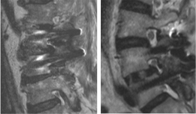
The study investigated whether the use of carbon fiber-reinforced PEEK screw material (CF-PEEK) can reduce magnetic resonance imaging (MRI) artifact formation. Two consecutive groups of patients were treated for degenerative spinal disorders of the lumbar spine with dorsal transpedicular spinal fusion. The first group (n = 27) received titanium pedicle screws. The second group (n = 20) received CF-PEEK screws. All patients underwent an MRI assessment within the first four postoperative weeks. For each operated segment, the surface of the artifact-free vertebral body area was calculated as percentage of the total vertebral body. For each implanted segment, the assessability of the spinal canal, the neuroforamina, and the pedicle screws, as well as the surrounding bony and soft-tissue structures was graded from 1 to 5. A mean artifact-free vertebral body area of 48.3 ± 5.0% was found in the in the titanium group and of 67.1 ± 5.6% in the CF-PEEK group (p ≤ 0.01). Assessability of the lumbar spine was significantly improved for CF-PEEK screws (p ≤ 0.01) for all measurements. CF-PEEK pedicle screws exhibit smaller artifact areas on vertebral body surfaces and their surrounding tissues, which improves the radiographic assessability. Hence, CF-PEEK may provide a diagnostic benefit.
本研究旨在探讨碳纤维增强型聚醚醚酮(CF-PEEK)螺钉材料的使用是否可以减少磁共振成像(MRI)伪影的形成。连续两组患有退行性腰椎疾病的患者接受了后路经椎弓根脊柱融合术。第一组(n=27)接受了钛制椎弓根螺钉治疗。第二组(n=20)接受了 CF-PEEK 螺钉治疗。所有患者均在术后 4 周内接受 MRI 评估。对于每个手术节段,计算无伪影的椎体区域面积占总椎体面积的百分比。对于每个植入节段,对椎管、神经孔、椎弓根螺钉以及周围的骨和软组织结构的可评估性进行 1 到 5 级评分。钛制组的无伪影椎体区域平均为 48.3±5.0%,CF-PEEK 组为 67.1±5.6%(p≤0.01)。对于所有测量值,CF-PEEK 螺钉的腰椎可评估性均显著提高(p≤0.01)。CF-PEEK 椎弓根螺钉在椎体表面及其周围组织上产生的伪影区域较小,这提高了影像学的可评估性。因此,CF-PEEK 可能具有诊断优势。