Kelly John P, Baran Francine M, Phillips James O, Weiss Avery H
Roger H. Johnson Vision Clinic, Seattle Children's Hospital, Division of Ophthalmology, Seattle, WA, USA.
University of Washington, Department of Ophthalmology, Seattle, WA, USA.
Transl Vis Sci Technol. 2020 Sep 22;9(10):21. doi: 10.1167/tvst.9.10.21. eCollection 2020 Sep.
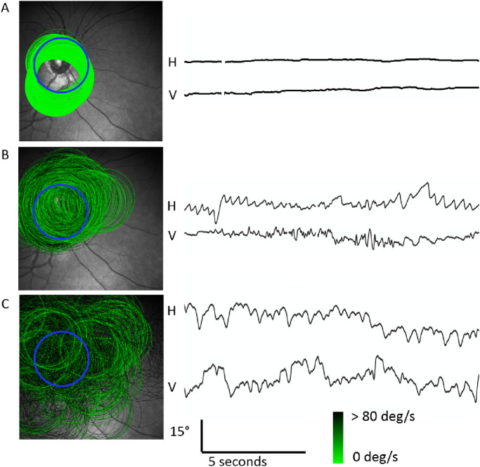
Poor fixation or nystagmus in children causes misalignment errors when measuring circumpapillary retinal nerve fiber layer (cpRNFL) thickness by simultaneous scanning laser ophthalmoscope imaging/optical coherence tomography (SLO/OCT). We investigated a method to assess cpRNFL from misaligned SLO/OCT scans.
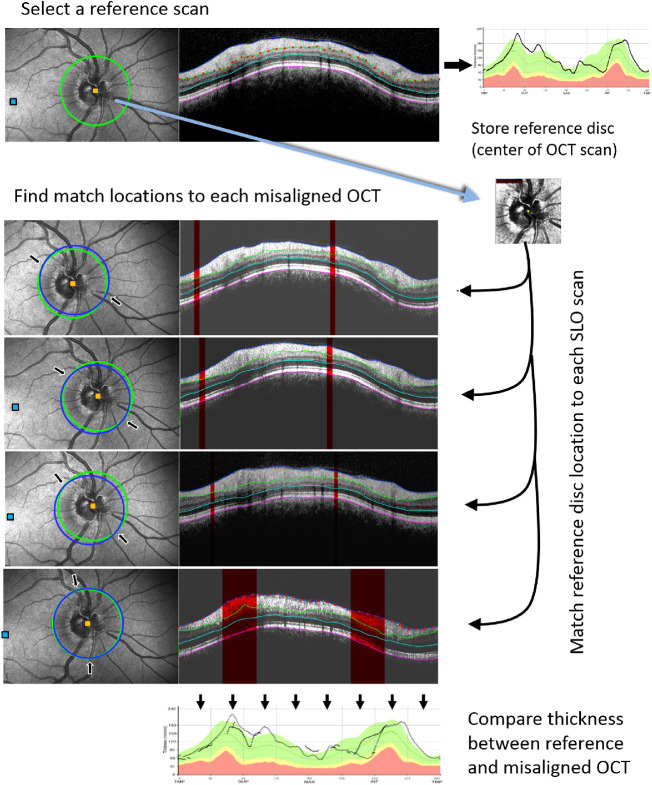
Heidelberg Spectralis SLO/OCT scans from a single clinical examination were retrospectively analyzed when automated eye tracking was unreliable. Retinal layer thickness was measured at overlapping match locations between a reference and misaligned scans based on the position data from simultaneously acquired SLO images. Three layers were segmented: cpRNFL, internal limiting membrane to outer nuclear layer (ILM-ONL), and total retinal thickness (TR). Accuracy was defined as the difference in thickness between the reference and misaligned scans at their match locations after correction for scan angle.
Thirty-five subjects, evaluated for glaucomatous nerve loss, met inclusion criteria. Group-averaged accuracy was -2.7, 1.4, and 0.3 µm for cpRNFL, ILM-ONL, and TR thickness, respectively. Across all layers, interobserver intraclass correlation coefficients ranged from 0.97 to 0.63 and the maximum Bland-Altman 95% limits of agreement were -21.6 to 20.7 µm. Variability was greatest for cpRNFL thickness and least for TR thickness. Increased variability was associated with lower signal-to-noise ratio but not with image-motion indices of shear, rotation, and scale.
Retinal layer thickness can be compared to a reference cpRNFL OCT scan when poor fixation and nystagmus causes misalignment errors. The analysis can be performed post hoc using multiple misaligned scans from standard SLO/OCT protocols.
Our method allows for assessment of cpRNFL in children who fail eye tracking.
儿童固定不佳或眼球震颤会在通过同步扫描激光检眼镜成像/光学相干断层扫描(SLO/OCT)测量视乳头周围视网膜神经纤维层(cpRNFL)厚度时导致对准误差。我们研究了一种从不对准的SLO/OCT扫描中评估cpRNFL的方法。
当自动眼跟踪不可靠时,对来自单次临床检查的海德堡Spectralis SLO/OCT扫描进行回顾性分析。基于同时采集的SLO图像的位置数据,在参考扫描和未对准扫描的重叠匹配位置测量视网膜层厚度。分割出三层:cpRNFL、内界膜至外核层(ILM-ONL)和视网膜总厚度(TR)。准确性定义为在对扫描角度进行校正后,参考扫描和未对准扫描在其匹配位置处的厚度差异。
35名接受青光眼性神经损伤评估的受试者符合纳入标准。cpRNFL、ILM-ONL和TR厚度的组平均准确性分别为-2.7、1.4和0.3μm。在所有层中,观察者间组内相关系数范围为0.97至0.63,最大Bland-Altman 95%一致性界限为-21.6至20.7μm。cpRNFL厚度的变异性最大,TR厚度的变异性最小。变异性增加与较低的信噪比相关,但与剪切、旋转和缩放的图像运动指数无关。
当固定不佳和眼球震颤导致对准误差时,视网膜层厚度可与参考cpRNFL OCT扫描进行比较。该分析可在事后使用来自标准SLO/OCT协议的多个未对准扫描进行。
我们的方法允许对眼跟踪失败的儿童进行cpRNFL评估。