Department of Applied Pharmacology, Kagoshima, Japan; Restorative Dentistry and Endodontology, Graduate School of Medical and Dental Sciences, Kagoshima University, Kagoshima, Japan.
Department of Applied Pharmacology, Kagoshima, Japan.
Free Radic Biol Med. 2020 Dec;161:60-70. doi: 10.1016/j.freeradbiomed.2020.09.027. Epub 2020 Oct 2.
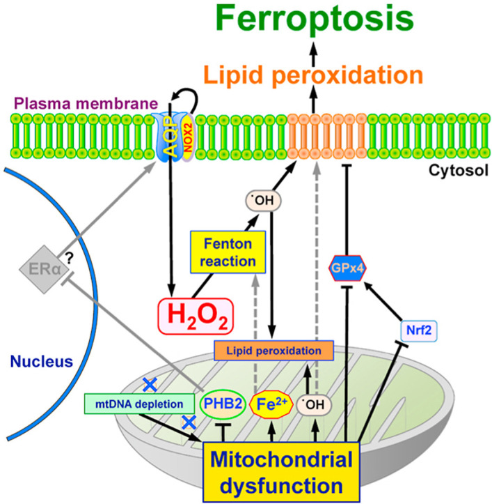
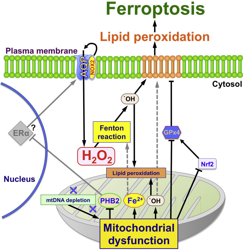
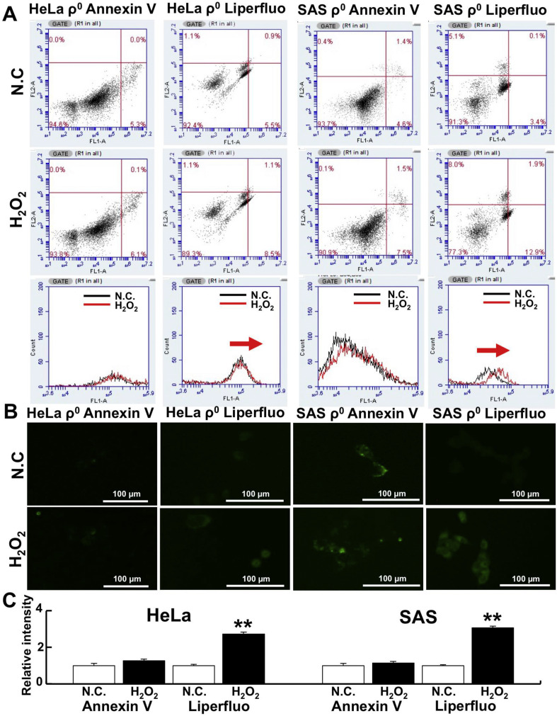
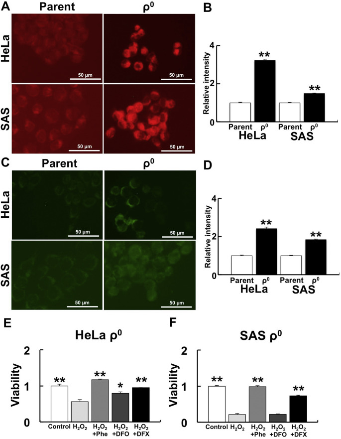
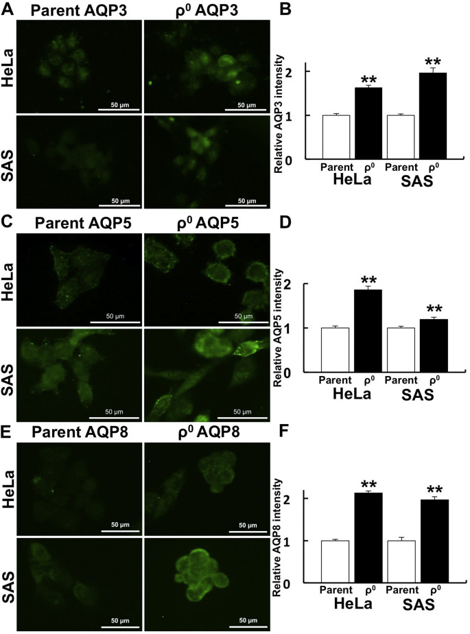
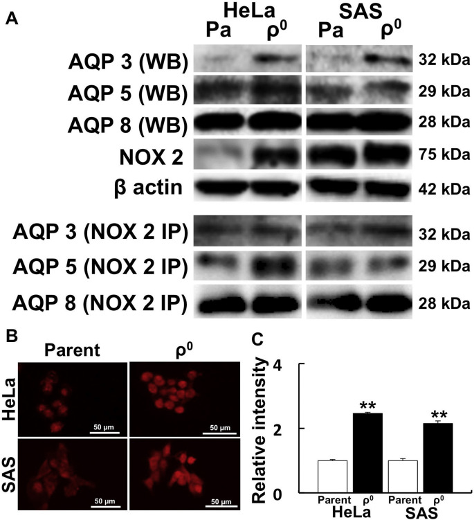
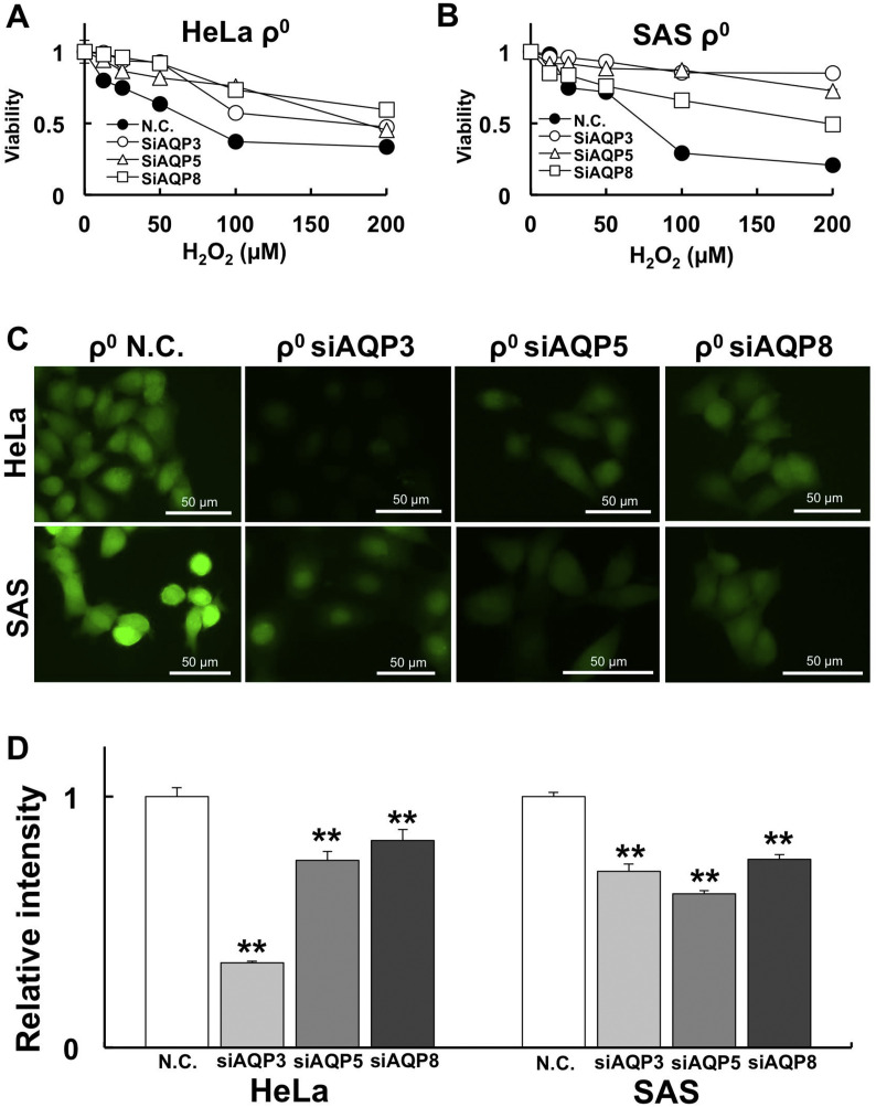
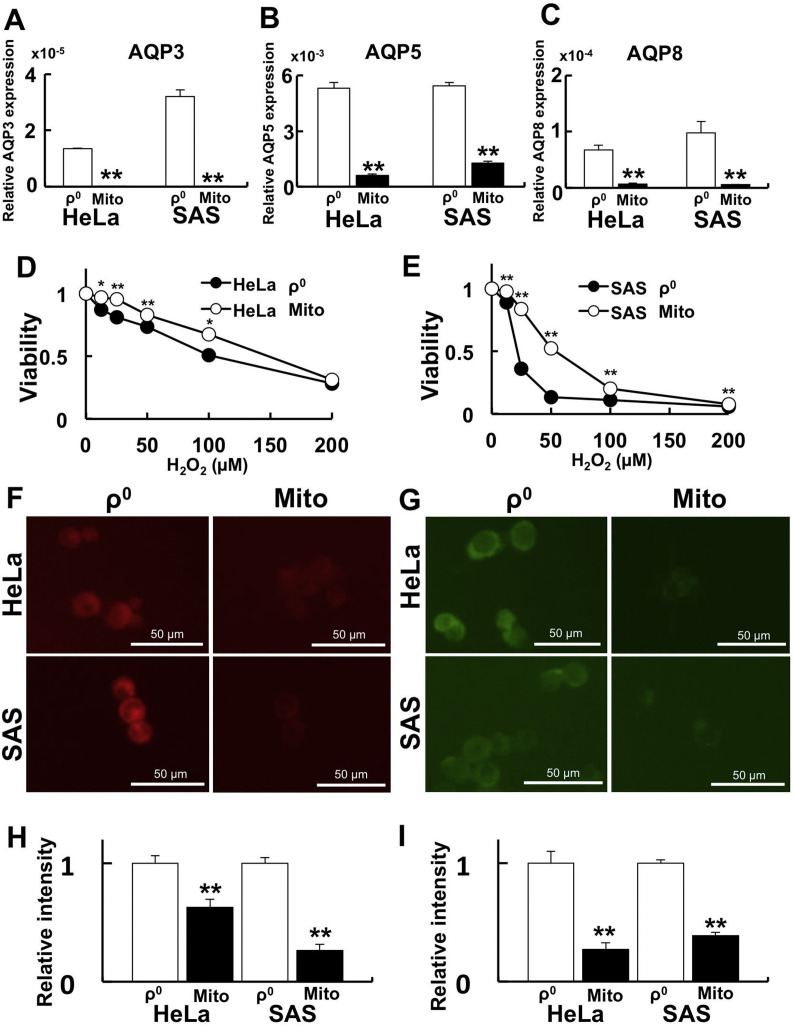
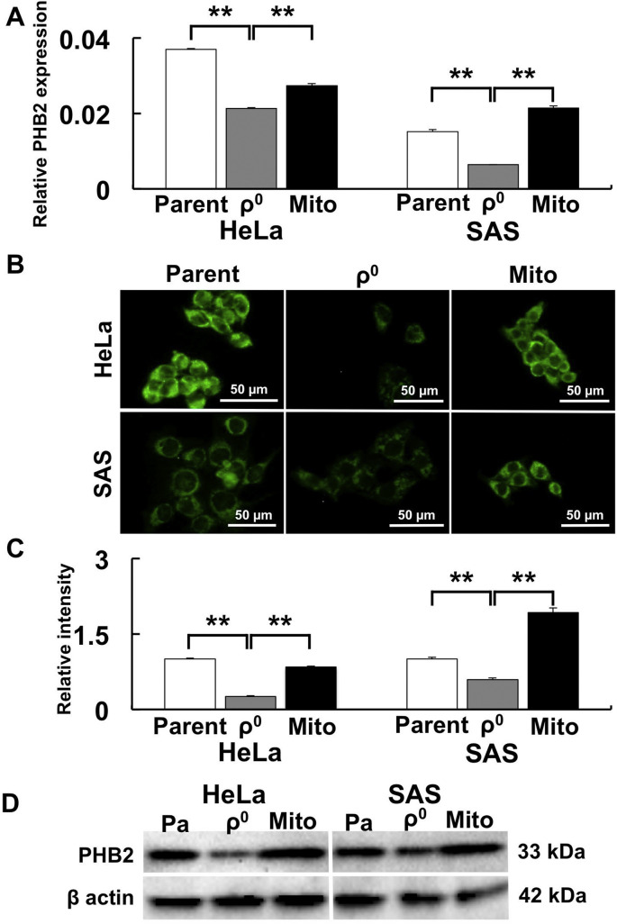
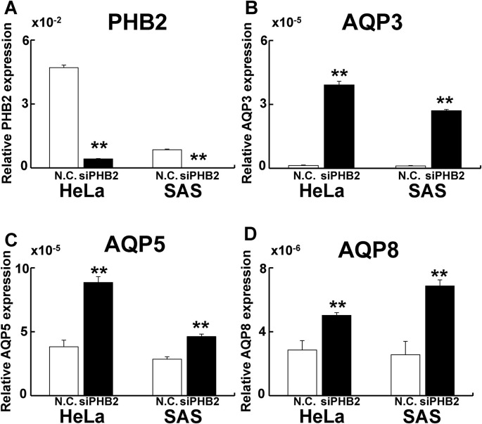
Most anti-cancer agents and radiotherapy exert their therapeutic effects via the production of free radicals. Ferroptosis is a recently described cell death process that is accompanied by iron-dependent lipid peroxidation. Hydrogen peroxide (HO) has been reported to induce cell death. However, it remains controversial whether HO-induced cell death is ferroptosis. In the present study, we aimed to elucidate the involvement of mitochondria in HO-induced ferroptosis and examined the molecules that regulate ferroptosis. We found that one mechanism underlying HO-induced cell death is ferroptosis, which occurs soon after HO treatment (within 3 h after HO treatment). We also investigated the involvement of mitochondria in HO-induced ferroptosis using mitochondrial DNA-depleted ρ cells because ρ cells produce more lipid peroxidation, hydroxyl radicals (OH), and are more sensitive to HO treatment. We found that ρ cells contain high Fe levels that lead to OH production by HO. Further, we observed that aquaporin (AQP) 3, 5, and 8 bind nicotinamide-adenine dinucleotide phosphate oxidase 2 and regulate the permeability of extracellular HO, thereby contributing to ferroptosis. Additionally, the role of mitochondria in ferroptosis was investigated using mitochondrial transfer in ρ cells. When mitochondria were transferred into ρ cells, the cells exhibited no sensitivity to HO-induced cytotoxicity because of decreased Fe levels. Moreover, mitochondrial transfer upregulated the mitochondrial quality control protein prohibitin 2 (PHB2), which contributes to reduced AQP expression. Our findings also revealed the involvement of AQP and PHB2 in ferroptosis. Our results indicate that HO treatment enhances AQP expression, Fe level, and lipid peroxidation, and decrease mitochondrial function by downregulating PHB2, and thus, is a promising modality for effective cancer treatment.
大多数抗癌药物和放射疗法通过产生自由基发挥治疗作用。铁死亡是一种新描述的细胞死亡过程,伴随着铁依赖性脂质过氧化。过氧化氢 (HO) 已被报道可诱导细胞死亡。然而,HO 诱导的细胞死亡是否是铁死亡仍存在争议。在本研究中,我们旨在阐明线粒体在 HO 诱导的铁死亡中的作用,并研究调节铁死亡的分子。我们发现,HO 诱导细胞死亡的一种机制是铁死亡,它发生在 HO 处理后不久(HO 处理后 3 小时内)。我们还使用线粒体 DNA 耗尽的 ρ 细胞研究了线粒体在 HO 诱导的铁死亡中的作用,因为 ρ 细胞产生更多的脂质过氧化、羟基自由基 (OH),并且对 HO 处理更敏感。我们发现 ρ 细胞中含有高浓度的 Fe,导致 HO 产生 OH。此外,我们观察到水通道蛋白 (AQP) 3、5 和 8 与烟酰胺腺嘌呤二核苷酸磷酸氧化酶 2 结合并调节细胞外 HO 的通透性,从而促进铁死亡。此外,还通过 ρ 细胞中的线粒体转移研究了线粒体在铁死亡中的作用。当线粒体被转移到 ρ 细胞中时,由于 Fe 水平降低,细胞对 HO 诱导的细胞毒性没有敏感性。此外,线粒体转移上调了线粒体质量控制蛋白 PHB2,这有助于减少 AQP 的表达。我们的研究结果还揭示了 AQP 和 PHB2 在铁死亡中的作用。我们的研究结果表明,HO 处理通过下调 PHB2 增强 AQP 表达、Fe 水平和脂质过氧化,降低线粒体功能,因此是一种有前途的有效癌症治疗方法。