Singapore Eye Research Institute, Singapore National Eye Centre, 11 Third Hospital Ave, Singapore, 168751, Singapore.
Ophthalmology and Visual Sciences Academic Clinical Program (Eye ACP), Duke-NUS Medical School, Singapore, Singapore.
Sci Rep. 2020 Oct 5;10(1):16505. doi: 10.1038/s41598-020-73585-0.
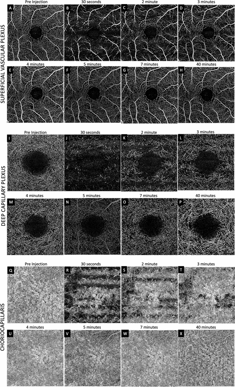
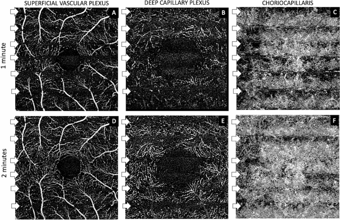
To describe patterns of reperfusion in the superficial vascular plexus (SVP), deep capillary plexus (DCP) and choriocapillaris (CC) as detected on optical coherence tomography (OCTA) in cynomogulus macaque monkey model following increase in intraocular pressure by an intravitreal injection. Animal imaging study. Two cynomogulus macaque monkeys. A 100 µL intravitreal injection (IVI) of saline was given in one eye of each monkey. Serial OCTA using a Zeiss Plex Elite 9000 was used to evaluate reperfusion patterns within the SCP, DCP, and CC. OCTA evidence of perfusion. Pulsation of the central retinal artery was detected after the intraocular pressure was elevated to 98 and ≥ 99 mmHg from IVI. Episodic flow within the SVP arterioles and venules and poor visualization of flow in capillaries was noted during the initial phase of elevated pressure. As the pressure declined, the flow signal within the DCP appeared initially as dots, which progressed laterally to loops which form capillary vortex configuration. Recovery of flow within the SVP and CC appeared sooner than in the DCP. At 40 min after the injection, well after the intraocular pressure normalized, the retinal and choriocapillaris vascular perfusion showed focal defects in every layer. Compared with pre-injection images, vessel density in the DCP was 68.8% and 78.6% of baseline in monkey 1 and monkey 2, respectively. In contrast vessel density in the SVP recovered to 84.2% and 88.9% of baseline. Increases in intraocular pressure from IVI have the potential to affect every layer of blood flow in the fundus. After nominal return of intraocular pressure, focal defects in flow persisted, which may result in longer term damage to the retina.
描述在眼内压升高时通过玻璃体内注射引起的猴模型中光学相干断层扫描血管造影(OCTA)检测到的浅层血管丛(SVP)、深层毛细血管丛(DCP)和脉络膜毛细血管(CC)的再灌注模式。动物影像学研究。两只食蟹猴。每只猴子的一只眼内注射 100µL 盐水。使用蔡司 Plex Elite 9000 进行连续 OCTA,以评估 SCP、DCP 和 CC 内的再灌注模式。OCTA 灌注证据。在眼内压从 IVI 升高到 98mmHg 和≥99mmHg 后,检测到中央视网膜动脉的搏动。在压力升高的初始阶段,注意到 SVP 动静脉中的间歇性血流和毛细血管中血流的不良可视化。随着压力下降,DCP 中的血流信号最初表现为点状,随后向环行侧向扩展,形成毛细血管涡流构型。SVP 和 CC 内的血流恢复比 DCP 更快。在注射后 40 分钟,即眼内压正常化后很久,视网膜和脉络膜毛细血管的血管灌注在每一层都显示出局灶性缺陷。与注射前图像相比,DCP 中的血管密度在猴 1 和猴 2 中分别为基线的 68.8%和 78.6%。相比之下,SVP 中的血管密度恢复到基线的 84.2%和 88.9%。从 IVI 增加眼内压有可能影响眼底各层的血流。在眼内压正常化后,血流的局灶性缺陷仍然存在,这可能导致视网膜的长期损伤。