Department of Ophthalmology, Korea University Guro Hospital, 148, Gurodong-ro, Guro-gu, Seoul, 08308, Republic of Korea.
Department of Ophthalmology, Hanoi Medical University, Hanoi, Vietnam.
Sci Rep. 2021 Apr 20;11(1):8555. doi: 10.1038/s41598-021-87689-8.
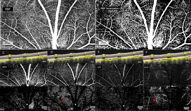
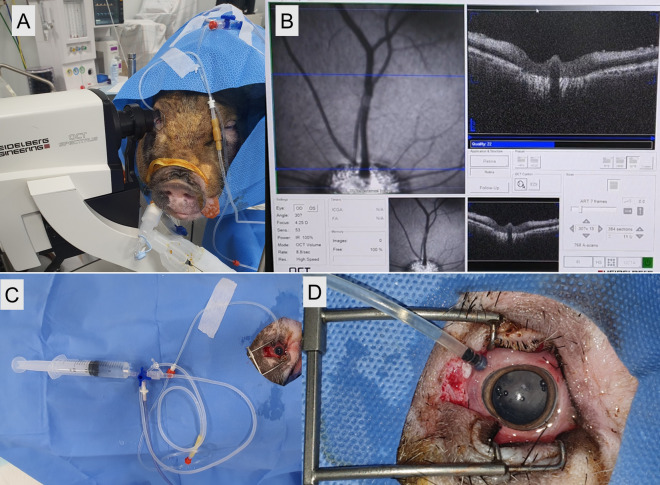
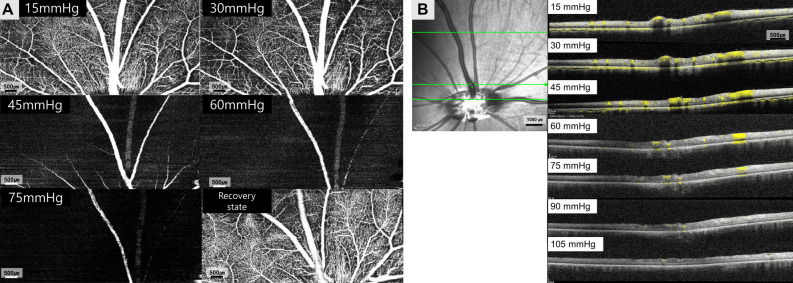
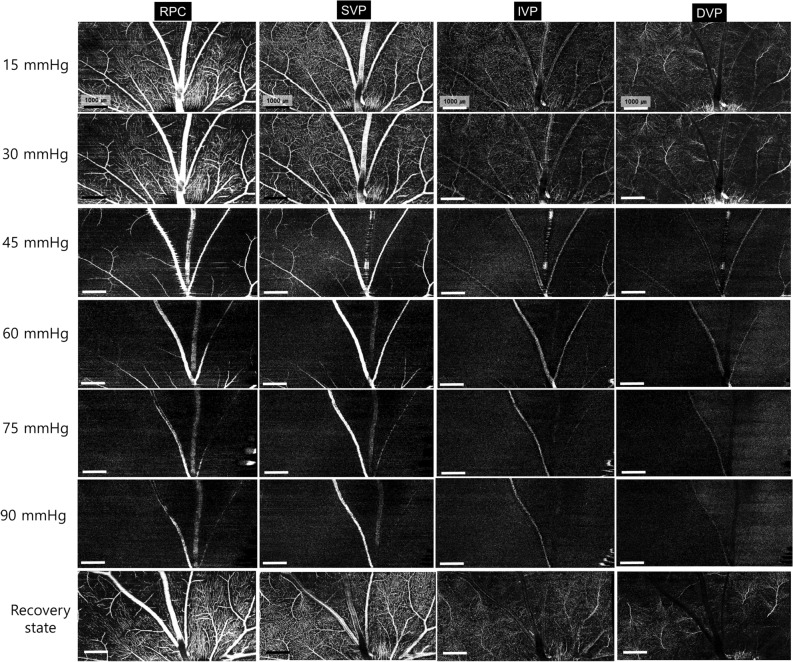
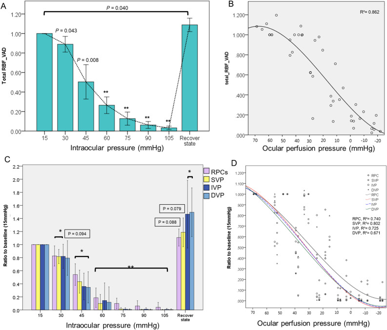
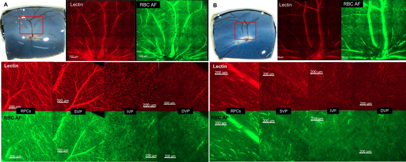
The purpose of this study was to evaluate density change in the retinal capillary plexus during intra ocular pressure (IOP) elevation in vitrectomized pigs' eyes using optical coherence tomography angiography (OCTA). Eight eyes of eight micro pigs received vitrectomy and the IOP was controlled from 15 mmHg (baseline) to 105 mmHg in 15 mmHg increments using a vented-gas forced-infusion system, and then decreased back to normal IOP (recovery state). The spectral-domain OCTA device was set to scan an area of 8.8 × 4.4 mm (30° × 15°) above the optic nerve head for each IOP. The relative vessel density (rVAD) compared to baseline was determined for the total retinal blood flow (RBF) which included major retinal artery and venous vessels, radial peripapillary capillaries (RPCs), superficial (SVP), intermediate (IVP), and deep vascular plexus (DVP). The mean rVAD was 0.890 in RBF, 0.826 in RPCs, 0.817 in SVP, 0.819 in IVP, and 0.794 in DVP at 30 mmHg. While the rVAD of RBF and RPCs decreased to 0.504 and 0.541 at 45 mmHg, the SVP, IVP, and DVP decreased to 0.433, 0.359, and 0.345, respectively. When IOP was normalized, the rVAD was recovered in all layers and the VAD of RBF, IVP, and DVP were higher than baseline (P = 0.040, 0.019, and 0.019, respectively). Retinal capillary density deterioration in each layer was found from 30 mmHg using an OCTA system which showed excellent depth-resolved segmentation of retinal capillary layers even at higher IOPs. Reduction in VAD showed full recovery after IOP normalization.
本研究旨在利用光相干断层扫描血管造影术(OCTA)评估玻璃体切割猪眼眼压(IOP)升高期间视网膜毛细血管丛的密度变化。8 只微型猪的 8 只眼接受玻璃体切割术,使用带通风孔的气体强制灌注系统将 IOP 从 15mmHg(基线)控制升高至 105mmHg,每 15mmHg 增加一次,然后降低至正常 IOP(恢复状态)。频谱 OCTA 设备设置为以视神经头上方 8.8×4.4mm(30°×15°)的区域进行扫描。对于包括主要视网膜动脉和静脉血管、放射状视神经乳头毛细血管(RPCs)、浅层(SVP)、中间(IVP)和深层血管丛(DVP)在内的总视网膜血流(RBF),与基线相比确定相对血管密度(rVAD)。在 30mmHg 时,RBF 的平均 rVAD 为 0.890,RPCs 为 0.826,SVP 为 0.817,IVP 为 0.819,DVP 为 0.794。当 IOP 升高至 45mmHg 时,RBF 和 RPCs 的 rVAD 降至 0.504 和 0.541,而 SVP、IVP 和 DVP 分别降至 0.433、0.359 和 0.345。当 IOP 正常化时,所有层的 rVAD 均得到恢复,RBF、IVP 和 DVP 的 VAD 高于基线(P=0.040、0.019 和 0.019)。即使在更高的 IOP 下,OCTA 系统也能出色地对视网膜毛细血管层进行深度分辨分割,从 30mmHg 开始发现各层视网膜毛细血管密度恶化。在 IOP 正常化后,VAD 减少完全恢复。