Department of Neurosurgery, Keio University School of Medicine, 35 Shinanomachi, Shinjuku-ku, Tokyo, 160-8582, Japan.
Department of Radiology, Keio University School of Medicine, 35 Shinanomachi, Shinjuku-ku, Tokyo, 160-8582, Japan.
Sci Rep. 2020 Oct 6;10(1):16623. doi: 10.1038/s41598-020-73658-0.
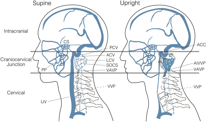
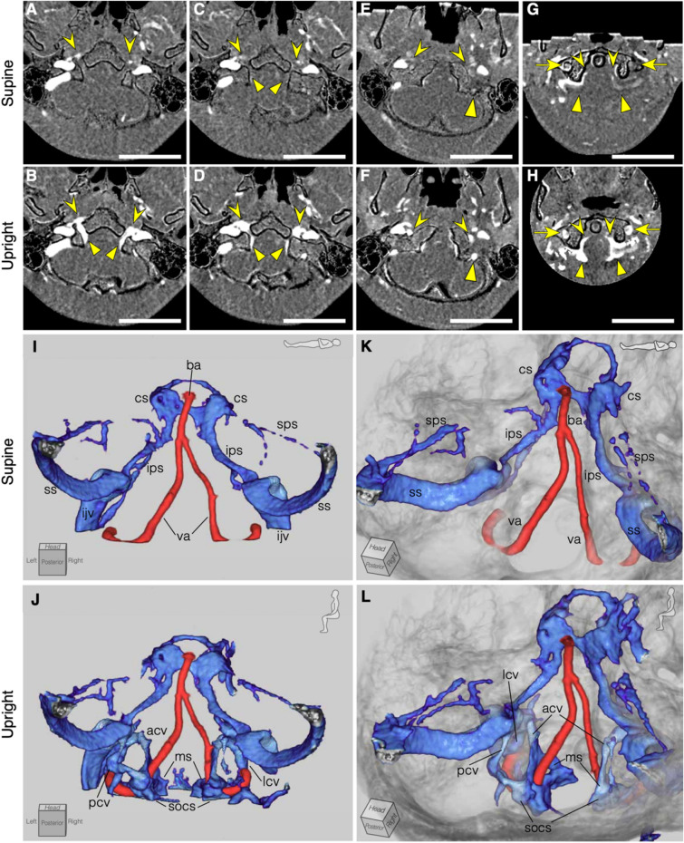
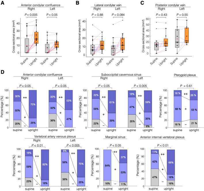
Since the venous system is affected by gravity, upright computed tomography (CT) in addition to conventional supine CT has great potential for evaluating postural changes in the venous system. We evaluated the morphological differences in the head and neck vessels by performing a contrast CT study in both the supine and the sitting positions. In this study, the 20 included participants (10 men and 10 women) were healthy adults aged 30 to 55 years. The cross-sectional area of the cervical vessels, craniocervical junction veins, and intracranial vessels were obtained quantitatively. Venous sinuses and venous plexuses that were difficult to measure were evaluated qualitatively. The average change in areas from a supine to an upright posture was - 77.87 ± 15.99% (P < 0.0001) in the right internal jugular vein (IJV), - 69.42 ± 23.15% (P < 0.0001) in the left IJV, - 61.52 ± 12.81% (P < 0.0001) in the right external jugular vein (EJV), and - 58.91 ± 17.37% (P < 0.0001) in the left EJV. In contrast, the change in the anterior condylar vein (ACV) from a supine to an upright posture was approximately + 144% (P < 0.005) on the right side and + 110% (P < 0.05) on the left side. In addition, according to the qualitative analysis, the posterior venous structures including the anterior condylar confluence (ACC) of the craniocervical junction became more prominent in an upright posture. Despite these changes, the intracranial vessels showed almost no change between postures. From a supine to an upright position, the IJVs and EJVs above the heart collapsed, and venous channels including the ACCs and ACVs opened, switching the main cerebral venous drainage from the IJVs to the vertebral venous system. Upright head CT angiography can be useful for investigating physiological and pathophysiological hemodynamics of the venous system accompanying postural changes.
由于静脉系统受重力影响,除了传统的仰卧位 CT 外,直立位 CT 对评估静脉系统的姿势变化具有很大的潜力。我们通过在仰卧位和坐位进行对比 CT 研究,评估头颈部血管的形态差异。在这项研究中,20 名纳入的参与者(10 名男性和 10 名女性)均为 30 至 55 岁的健康成年人。定量获得了颈部血管、颅颈交界处静脉和颅内血管的横截面积。定性评估了难以测量的静脉窦和静脉丛。从仰卧位到直立位,右侧颈内静脉(IJV)的面积平均变化为-77.87±15.99%(P<0.0001),左侧 IJV 为-69.42±23.15%(P<0.0001),右侧颈外静脉(EJV)为-61.52±12.81%(P<0.0001),左侧 EJV 为-58.91±17.37%(P<0.0001)。相比之下,右侧前髁静脉(ACV)从仰卧位到直立位的变化约为+144%(P<0.005),左侧为+110%(P<0.05)。此外,根据定性分析,颅颈交界处的前髁汇合处(ACC)等后向静脉结构在直立位时变得更加突出。尽管存在这些变化,但颅内血管在体位之间几乎没有变化。从仰卧位到直立位,心脏上方的 IJVs 和 EJVs 塌陷,包括 ACCs 和 ACVs 的静脉通道打开,将大脑主要静脉引流从 IJVs 切换到椎静脉系统。直立头 CT 血管造影术可用于研究伴随姿势变化的静脉系统的生理和病理生理学血流动力学。