Department of Neurosurgery, TUM Neuroimaging Center, Technical University of Munich, Germany, School of Medicine, Klinikum rechts der Isar, Ismaninger Str. 22, 81675, Munich, Germany.
Centre for Language and Cognition Groningen (CLCG), University of Groningen, Groningen, the Netherlands.
Acta Neurochir (Wien). 2021 Apr;163(4):895-903. doi: 10.1007/s00701-020-04545-w. Epub 2020 Oct 7.
The human white matter pathway network is complex and of critical importance for functionality. Thus, learning and understanding white matter tract anatomy is important for the training of neuroscientists and neurosurgeons. The study aims to test and evaluate a new method for fiber dissection using augmented reality (AR) in a group which is experienced in cadaver white matter dissection courses and in vivo tractography.
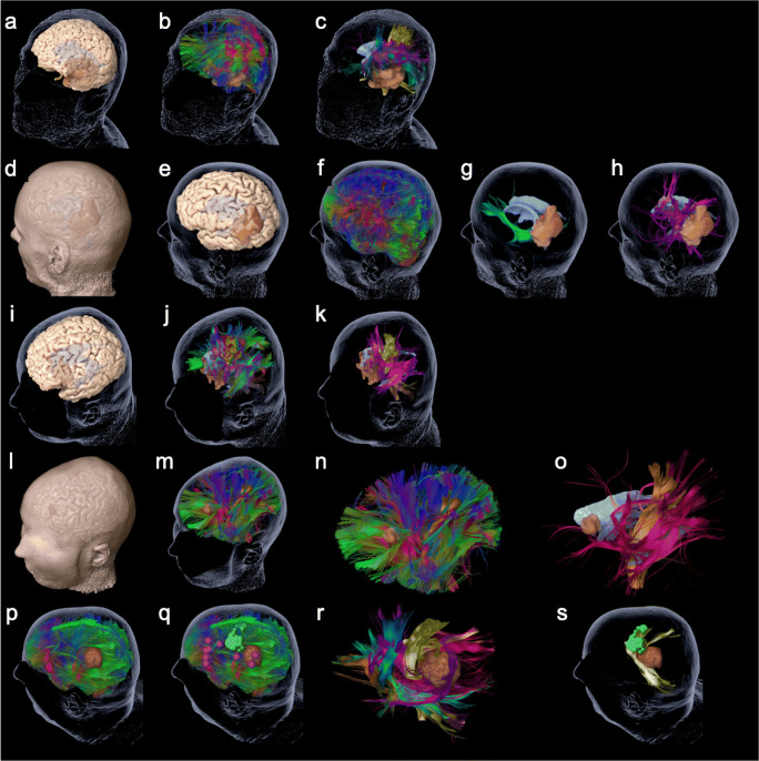
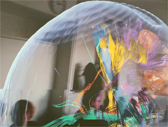
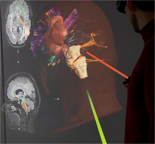
Fifteen neurosurgeons, neurolinguists, and neuroscientists participated in this questionnaire-based study. We presented five cases of patients with left-sided perisylvian gliomas who underwent awake craniotomy. Diffusion tensor imaging fiber tracking (DTI FT) was performed and the language-related networks were visualized separated in different tracts by color. Participants were able to virtually dissect the prepared DTI FTs using a spatial computer and AR goggles. The application was evaluated through a questionnaire with answers from 0 (minimum) to 10 (maximum).
Participants rated the overall experience of AR fiber dissection with a median of 8 points (mean ± standard deviation 8.5 ± 1.4). Usefulness for fiber dissection courses and education in general was rated with 8 (8.3 ± 1.4) and 8 (8.1 ± 1.5) points, respectively. Educational value was expected to be high for several target audiences (student: median 9, 8.6 ± 1.4; resident: 9, 8.5 ± 1.8; surgeon: 9, 8.2 ± 2.4; scientist: 8.5, 8.0 ± 2.4). Even clinical application of AR fiber dissection was expected to be of value with a median of 7 points (7.0 ± 2.5).
The present evaluation of this first application of AR for fiber dissection shows a throughout positive evaluation for educational purposes.
人类白质通路网络复杂,对功能至关重要。因此,学习和理解白质束解剖结构对于神经科学家和神经外科医生的培训很重要。本研究旨在测试和评估一种新的方法,即在有尸体白质解剖课程和活体束追踪经验的小组中使用增强现实(AR)进行纤维解剖。
15 名神经外科医生、神经语言学家和神经科学家参加了这项基于问卷的研究。我们展示了 5 例接受清醒开颅术的左侧大脑外侧裂胶质瘤患者的病例。进行弥散张量成像纤维追踪(DTI FT),并通过颜色将与语言相关的网络可视化分离到不同的束中。参与者可以使用空间计算机和 AR 护目镜虚拟解剖准备好的 DTI FT。通过回答从 0(最低)到 10(最高)的问卷评估应用程序。
参与者对 AR 纤维解剖的总体体验评分中位数为 8 分(平均值±标准差 8.5±1.4)。纤维解剖课程和一般教育的有用性评分分别为 8(8.3±1.4)和 8(8.1±1.5)。几个目标受众预计具有很高的教育价值(学生:中位数 9,8.6±1.4;住院医师:9,8.5±1.8;外科医生:9,8.2±2.4;科学家:8.5,8.0±2.4)。甚至预计 AR 纤维解剖的临床应用也具有价值,中位数为 7 分(7.0±2.5)。
本研究首次应用 AR 进行纤维解剖的评估结果表明,其具有积极的教育意义。