Kirilina Evgeniya, Helbling Saskia, Morawski Markus, Pine Kerrin, Reimann Katja, Jankuhn Steffen, Dinse Juliane, Deistung Andreas, Reichenbach Jürgen R, Trampel Robert, Geyer Stefan, Müller Larissa, Jakubowski Norbert, Arendt Thomas, Bazin Pierre-Louis, Weiskopf Nikolaus
Department of Neurophysics, Max Planck Institute for Human Cognitive and Brain Sciences, Stephanstraße 1a, 04103 Leipzig, Germany.
Center for Cognitive Neuroscience Berlin, Free University Berlin, Habelschwerdter Allee 45, 14195 Berlin, Germany.
Sci Adv. 2020 Oct 7;6(41). doi: 10.1126/sciadv.aaz9281. Print 2020 Oct.
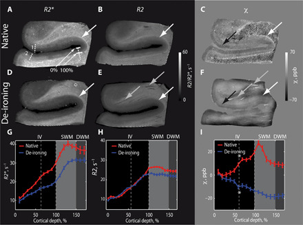
Superficial white matter (SWM) contains the most cortico-cortical white matter connections in the human brain encompassing the short U-shaped association fibers. Despite its importance for brain connectivity, very little is known about SWM in humans, mainly due to the lack of noninvasive imaging methods. Here, we lay the groundwork for systematic in vivo SWM mapping using ultrahigh resolution 7 T magnetic resonance imaging. Using biophysical modeling informed by quantitative ion beam microscopy on postmortem brain tissue, we demonstrate that MR contrast in SWM is driven by iron and can be linked to the microscopic iron distribution. Higher SWM iron concentrations were observed in U-fiber-rich frontal, temporal, and parietal areas, potentially reflecting high fiber density or late myelination in these areas. Our SWM mapping approach provides the foundation for systematic studies of interindividual differences, plasticity, and pathologies of this crucial structure for cortico-cortical connectivity in humans.
脑白质浅层(SWM)包含人脑皮质与皮质之间最多的白质连接,包括短U形联合纤维。尽管其对脑连接性很重要,但由于缺乏无创成像方法,人们对人类的SWM知之甚少。在此,我们利用超高分辨率7T磁共振成像为系统的活体SWM映射奠定基础。通过对死后脑组织进行定量离子束显微镜检查获得的生物物理模型,我们证明SWM中的磁共振对比由铁驱动,并且可以与微观铁分布相关联。在富含U纤维的额叶、颞叶和顶叶区域观察到较高的SWM铁浓度,这可能反映了这些区域的高纤维密度或晚期髓鞘形成。我们的SWM映射方法为系统研究人类皮质与皮质连接这一关键结构的个体差异、可塑性和病理学提供了基础。