PEIRENE, EA 7500, University of Limoges, 123 Avenue Albert Thomas, 87060, Limoges, France.
XLIM, UMR 7252, University of Limoges, 123 Avenue Albert Thomas, 87060, Limoges, France.
Sci Rep. 2020 Oct 7;10(1):16749. doi: 10.1038/s41598-020-74021-z.
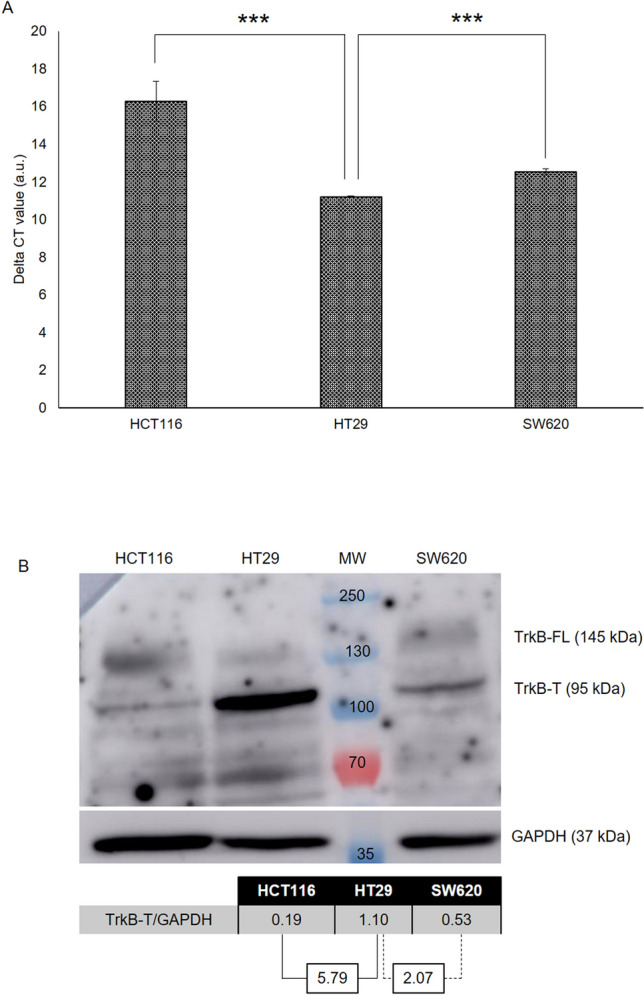
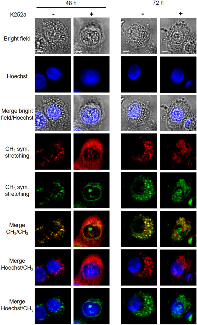
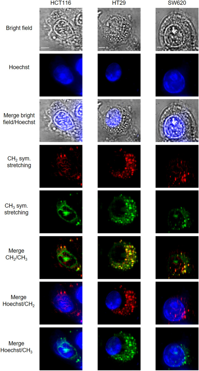
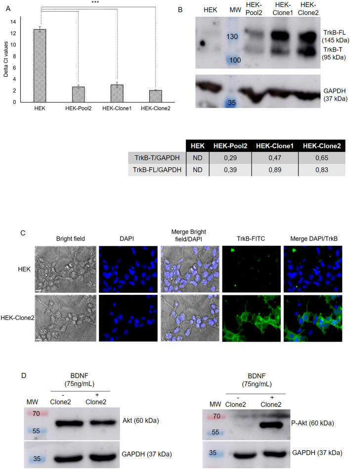
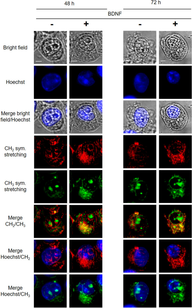
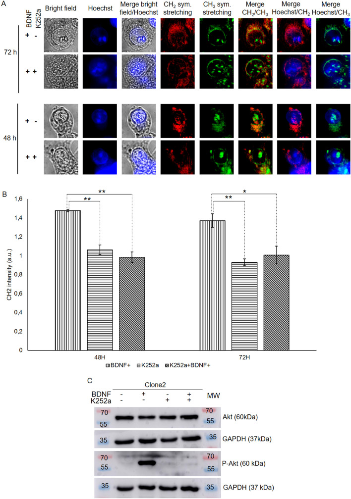
For many years, scientists have been looking for specific biomarkers associated with cancer cells for diagnosis purposes. These biomarkers mainly consist of proteins located at the cell surface (e.g. the TrkB receptor) whose activation is associated with specific metabolic modifications. Identification of these metabolic changes usually requires cell fixation and specific dye staining. MCARS microspectroscopy is a label-free, non-toxic, and minimally invasive method allowing to perform analyses of live cells and tissues. We used this method to follow the formation of lipid droplets in three colorectal cancer cell lines expressing TrkB. MCARS images of cells generated from signal integration of CH stretching modes allow to discriminate between lipid accumulation in the endoplasmic reticulum and the formation of cytoplasmic lipid droplets. We found that the number of the latter was related to the TrkB expression level. This result was confirmed thanks to the creation of a HEK cell line which over-expresses TrkB. We demonstrated that BDNF-induced TrkB activation leads to the formation of cytoplasmic lipid droplets, which can be abolished by K252a, an inhibitor of TrkB. So, MCARS microspectroscopy proved useful in characterizing cancer cells displaying an aberrant lipid metabolism.
多年来,科学家们一直在寻找与癌细胞相关的特定生物标志物,用于诊断目的。这些生物标志物主要由位于细胞表面的蛋白质组成(例如 TrkB 受体),其激活与特定的代谢修饰有关。这些代谢变化的识别通常需要细胞固定和特定的染料染色。MCARS 微光谱是一种无标记、无毒且微创的方法,可用于对活细胞和组织进行分析。我们使用这种方法来跟踪三种表达 TrkB 的结直肠癌细胞系中脂滴的形成。来自 CH 伸缩模式信号集成的细胞 MCARS 图像允许区分内质网中的脂质积累和细胞质脂滴的形成。我们发现后者的数量与 TrkB 的表达水平有关。这一结果通过创建过表达 TrkB 的 HEK 细胞系得到了证实。我们证明 BDNF 诱导的 TrkB 激活导致细胞质脂滴的形成,而 TrkB 的抑制剂 K252a 可以阻止这种形成。因此,MCARS 微光谱在表征显示异常脂质代谢的癌细胞方面非常有用。