Instituto Universitario de Tecnologías de la Información y Comunicaciones, Universitat Politècnica de València, València, Spain.
Department of Information Technology, Uppsala University, Uppsala, Sweden.
PLoS One. 2020 Oct 14;15(10):e0232500. doi: 10.1371/journal.pone.0232500. eCollection 2020.
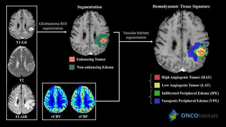
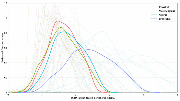
Genetic classifications are crucial for understanding the heterogeneity of glioblastoma. Recently, perfusion MRI techniques have demonstrated associations molecular alterations. In this work, we investigated whether perfusion markers within infiltrated peripheral edema were associated with proneural, mesenchymal, classical and neural subtypes.
ONCOhabitats open web services were used to obtain the cerebral blood volume at the infiltrated peripheral edema for MRI studies of 50 glioblastoma patients from The Cancer Imaging Archive: TCGA-GBM. ANOVA and Kruskal-Wallis tests were carried out in order to assess the association between vascular features and the Verhaak subtypes. For assessing specific differences, Mann-Whitney U-test was conducted. Finally, the association of overall survival with molecular and vascular features was assessed using univariate and multivariate Cox models.
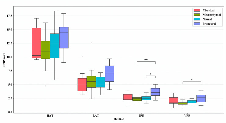
ANOVA and Kruskal-Wallis tests for the maximum cerebral blood volume at the infiltrated peripheral edema between the four subclasses yielded false discovery rate corrected p-values of <0.001 and 0.02, respectively. This vascular feature was significantly higher (p = 0.0043) in proneural patients compared to the rest of the subtypes while conducting Mann-Whitney U-test. The multivariate Cox model pointed to redundant information provided by vascular features at the peripheral edema and proneural subtype when analyzing overall survival.
Higher relative cerebral blood volume at infiltrated peripheral edema is associated with proneural glioblastoma subtype suggesting underlying vascular behavior related to molecular composition in that area.
遗传分类对于理解胶质母细胞瘤的异质性至关重要。最近,灌注 MRI 技术已经证明了与分子改变的相关性。在这项工作中,我们研究了浸润性周边水肿内的灌注标志物是否与前神经型、间质型、经典型和神经型亚型相关。
使用 ONCOhabitats 开放网络服务,我们从癌症影像档案库(TCGA-GBM)中获得了 50 名胶质母细胞瘤患者 MRI 研究的浸润性周边水肿脑血容量。进行方差分析和 Kruskal-Wallis 检验,以评估血管特征与 Verhaak 亚型之间的关联。为了评估特定差异,进行了 Mann-Whitney U 检验。最后,使用单变量和多变量 Cox 模型评估总生存期与分子和血管特征的关联。
对浸润性周边水肿的最大脑血容量进行方差分析和 Kruskal-Wallis 检验,在四个亚类之间的校正错误发现率 p 值分别为<0.001 和 0.02。在进行 Mann-Whitney U 检验时,这种血管特征在前神经型患者中明显更高(p = 0.0043)。多变量 Cox 模型指出,在分析总生存期时,周边水肿和前神经型的血管特征提供了冗余信息。
浸润性周边水肿的相对脑血容量较高与前神经胶质母细胞瘤亚型相关,提示该区域的血管行为与分子组成有关。