Lamberts A, Kotnik N, Diercks G F H, Meijer J M, Di Zenzo G, Pas H H, Jonkman M F, Gibbs B F, Raap U, Horváth B
Department of Dermatology, University Medical Center Groningen, University of Groningen, Groningen, the Netherlands.
Department of Experimental Dermatology and Allergology, University of Oldenburg, Oldenburg, Germany.
J Eur Acad Dermatol Venereol. 2021 Apr;35(4):973-980. doi: 10.1111/jdv.16996. Epub 2020 Nov 17.
Non-bullous pemphigoid (NBP) is a pemphigoid variant which frequently resembles other pruritic skin diseases. In contrast with bullous pemphigoid (BP), blisters are absent. In BP, previous studies showed that IgE autoantibodies may be involved in its pathogenesis. IgE-activated mast cells, basophils and eosinophils may participate in BP by inducing pruritus and possibly blister formation, although the differential role of IgE in NBP compared with BP has not yet been described.
To assess IgE in serum and skin of NBP and BP patients.
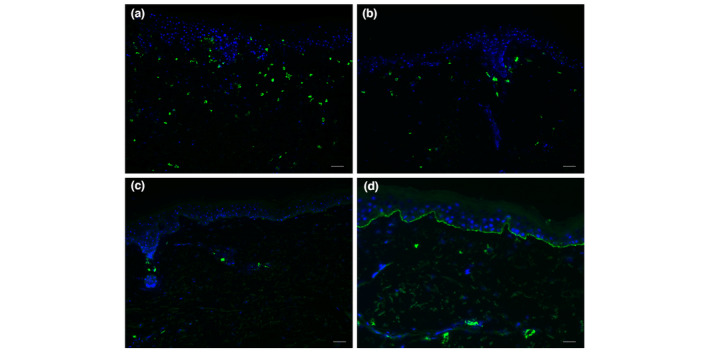
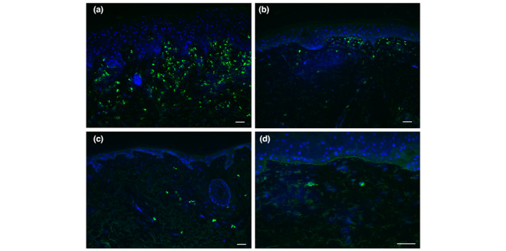
We examined total IgE and pemphigoid-specific IgE in the serum of 68 NBP and 50 BP patients by enzyme-linked immunosorbent assay (ELISA). Sera of 25 pemphigus patients and 25 elderly patients with pruritus were included as controls. Skin biopsies of 14 NBP and 14 BP patients with the highest IgE titres to NC16A were stained for IgE by immunofluorescence techniques.
Total IgE was elevated in 63% of NBP and 60% of BP patients, and in 20% of pemphigus controls, as well as 60% of elderly controls. IgE ELISAs were more frequently positive in BP than in NBP (NC16A 18% vs. 9%, P = 0.139; BP230 34% vs. 22%, P = 0.149). IgE ELISAs for NC16A and BP230 were positive in 8% and 20% of elderly controls, respectively, while all pemphigus controls were negative. Two of 28 biopsies (7%; one NBP, one BP) showed linear IgE along the basement membrane zone, while in most biopsies (71% NBP; 86% BP) IgE was bound to dermal cells.
Since IgE was present in the serum and skin of both NBP and BP patients, this supports IgE-dependent mechanisms common to both diseases, such as pruritus. However, it remains to be elucidated whether IgE contributes to blister formation in BP.
非大疱性类天疱疮(NBP)是类天疱疮的一种变体,常与其他瘙痒性皮肤病相似。与大疱性类天疱疮(BP)不同,NBP没有水疱。在BP中,先前的研究表明IgE自身抗体可能参与其发病机制。IgE激活的肥大细胞、嗜碱性粒细胞和嗜酸性粒细胞可能通过诱导瘙痒并可能参与水疱形成而参与BP的发病过程,尽管与BP相比,IgE在NBP中的不同作用尚未见报道。
评估NBP和BP患者血清及皮肤中的IgE。
我们采用酶联免疫吸附测定(ELISA)检测了68例NBP患者和50例BP患者血清中的总IgE和类天疱疮特异性IgE。纳入25例天疱疮患者和25例老年瘙痒患者的血清作为对照。对14例对NC16A的IgE滴度最高的NBP患者和14例BP患者的皮肤活检标本采用免疫荧光技术进行IgE染色。
63%的NBP患者、60%的BP患者、20%的天疱疮对照患者以及60%的老年对照患者的总IgE升高。IgE ELISA在BP患者中的阳性率高于NBP患者(NC16A分别为18%对9%,P = 0.139;BP230分别为34%对22%,P = 0.149)。老年对照患者中,针对NC16A和BP230的IgE ELISA阳性率分别为8%和20%,而所有天疱疮对照患者均为阴性。28例活检标本中有2例(7%;1例NBP,1例BP)在基底膜带显示线性IgE,而在大多数活检标本中(71%的NBP;86%的BP)IgE与真皮细胞结合。
由于IgE存在于NBP和BP患者的血清及皮肤中,这支持了两种疾病共有的IgE依赖性机制,如瘙痒。然而,IgE是否促成BP中的水疱形成仍有待阐明。