Barraso Marina, Alé-Chilet Aníbal, Hernández Teresa, Oliva Cristian, Vinagre Irene, Ortega Emilio, Figueras-Roca Marc, Sala-Puigdollers Anna, Esquinas Cristina, Esmatjes Enric, Adán Alfredo, Zarranz-Ventura Javier
Institut Clínic d'Oftalmologia (ICOF), Hospital Clínic, Barcelona, Spain.
Diabetes Unit, Institut Clínic de Malalties Digestives i Metabòliques (ICMDM), Hospital Clínic, Barcelona, Spain.
Transl Vis Sci Technol. 2020 Sep 30;9(10):34. doi: 10.1167/tvst.9.10.34. eCollection 2020 Sep.
The purpose of this study was to evaluate specifically in type 1 diabetes mellitus (DM) individuals the relationship between perifoveal superficial capillary plexus (SCP) parameters assessed by optical coherence tomography angiography (OCTA) and diabetic retinopathy (DR) grade.
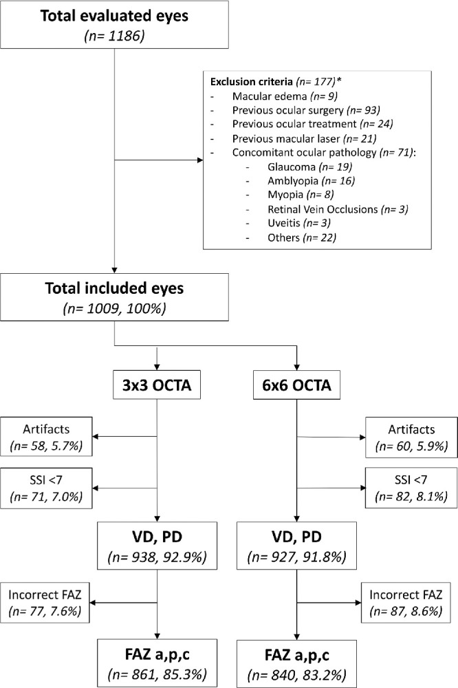
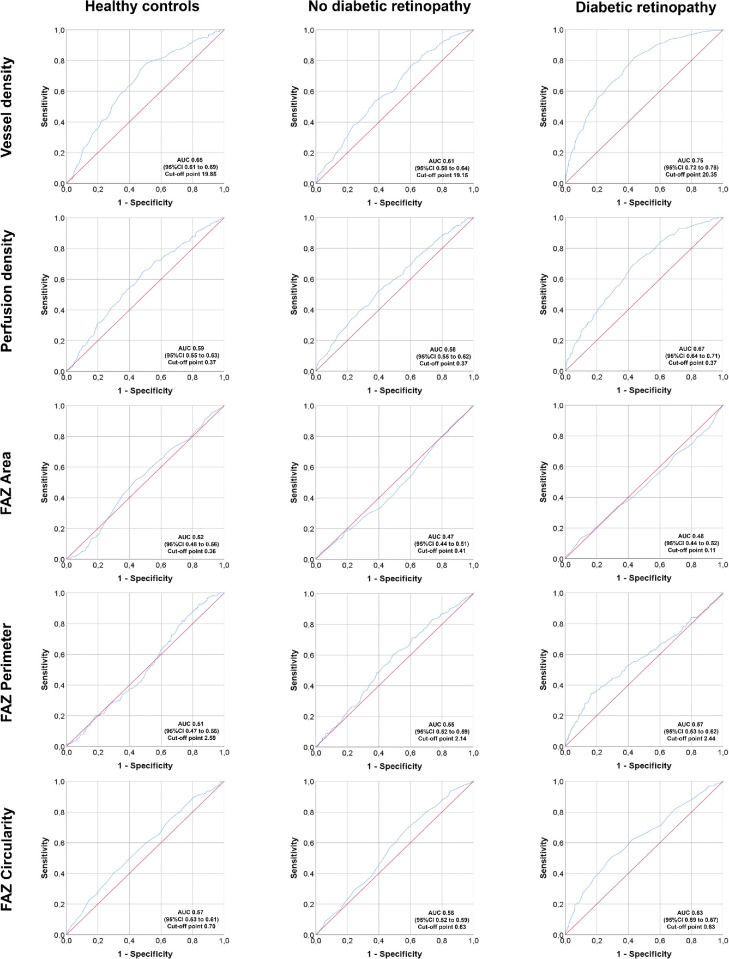
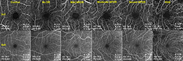
Cross-sectional analysis of a large scale prospective OCTA trial cohort (ClinicalTrials.gov NCT03422965). A total of 1186 eyes (593 individuals), 956 type 1 DM eyes (478 patients), and 230 control eyes (115 healthy volunteers) were included in this study. DR stage was graded according to the International Classification. OCTA imaging was performed with a commercially available device (Cirrus HD-OCT). Vessel density (VD), perfusion density (PD), and foveal avascular zone (FAZ) area, perimeter and circularity measurements were quantified in the SCP and receiver operating characteristic (ROC) curves were constructed for each OCTA parameter.
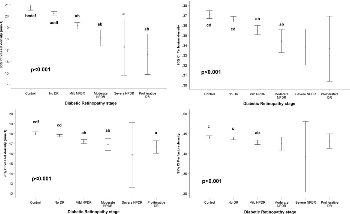
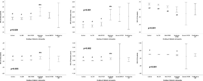
VD and PD (in both 3 × 3 and 6 × 6 mm captures) were inversely associated with DR stage ( < 0.001 in all cases) in a multiple regression analysis after controlling by age, gender, signal strength index, axial length, and DM duration. Greater FAZ area and perimeter and conversely lower circularity measurements were observed as DR severity increased in both scanning protocols ( < 0.05 in all cases).
In type 1 DM individuals, OCTA provides an objective, continuous, and reliable method for accurate quantification of VD, PD, and FAZ parameters in the SCP, which ultimately correlate with DR stages.
Objective OCTA measurements of the retinal microvasculature could substitute the clinical DR classification in patients with type 1 DM, identify patients at risk of DR progression, and inform treatment decisions to modify the evolution of the disease.
本研究旨在专门评估1型糖尿病(DM)患者中,通过光学相干断层扫描血管造影(OCTA)评估的黄斑周围浅表毛细血管丛(SCP)参数与糖尿病视网膜病变(DR)分级之间的关系。
对一项大规模前瞻性OCTA试验队列(ClinicalTrials.gov NCT03422965)进行横断面分析。本研究共纳入1186只眼(593名个体),其中956只1型糖尿病眼(478例患者)和230只对照眼(115名健康志愿者)。DR分期根据国际分类法进行分级。使用市售设备(Cirrus HD-OCT)进行OCTA成像。在SCP中量化血管密度(VD)、灌注密度(PD)以及黄斑无血管区(FAZ)的面积、周长和圆形度测量值,并为每个OCTA参数构建受试者操作特征(ROC)曲线。
在控制年龄、性别、信号强度指数、眼轴长度和糖尿病病程后进行的多元回归分析中,VD和PD(在3×3和6×6 mm采集区域中)均与DR分期呈负相关(所有情况均P<0.001)。在两种扫描方案中,随着DR严重程度增加,均观察到FAZ面积和周长增大,圆形度测量值降低(所有情况均P<0.05)。
在1型糖尿病患者中,OCTA为准确量化SCP中的VD、PD和FAZ参数提供了一种客观、连续且可靠的方法,这些参数最终与DR分期相关。
对视网膜微血管进行客观的OCTA测量可替代1型糖尿病患者的临床DR分级,识别有DR进展风险的患者,并为改变疾病进展的治疗决策提供依据。