Carrera-Escalé Laura, Benali Anass, Rathert Ann-Christin, Martín-Pinardel Ruben, Bernal-Morales Carolina, Alé-Chilet Anibal, Barraso Marina, Marín-Martinez Sara, Feu-Basilio Silvia, Rosinés-Fonoll Josep, Hernandez Teresa, Vilá Irene, Castro-Dominguez Rafael, Oliva Cristian, Vinagre Irene, Ortega Emilio, Gimenez Marga, Vellido Alfredo, Romero Enrique, Zarranz-Ventura Javier
Intelligent Data Science and Artificial Intelligence (IDEAI) Research Center.
Department of Computer Science, Facultat d'Informàtica de Barcelona (FIB), Universitat Politècnica de Catalunya (UPC), Barcelona, Spain.
Ophthalmol Sci. 2022 Nov 21;3(2):100259. doi: 10.1016/j.xops.2022.100259. eCollection 2023 Jun.
To evaluate the diagnostic accuracy of machine learning (ML) techniques applied to radiomic features extracted from OCT and OCT angiography (OCTA) images for diabetes mellitus (DM), diabetic retinopathy (DR), and referable DR (R-DR) diagnosis.
Cross-sectional analysis of a retinal image dataset from a previous prospective OCTA study (ClinicalTrials.govNCT03422965).
Patients with type 1 DM and controls included in the progenitor study.
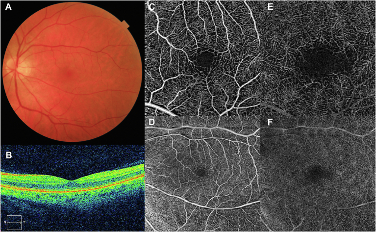
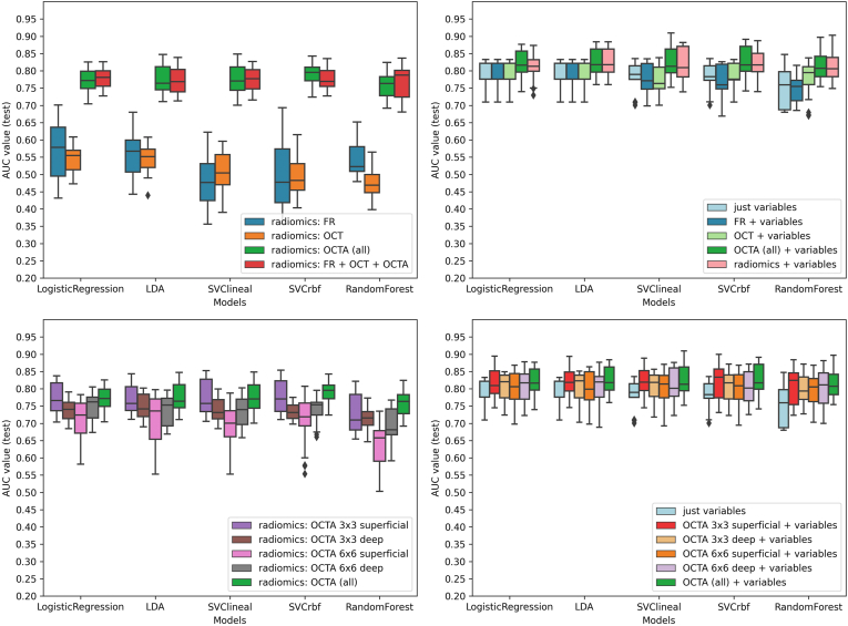
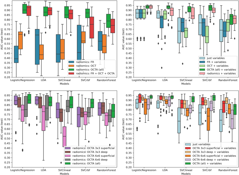
Radiomic features were extracted from fundus retinographies, OCT, and OCTA images in each study eye. Logistic regression, linear discriminant analysis, support vector classifier (SVC)-linear, SVC-radial basis function, and random forest models were created to evaluate their diagnostic accuracy for DM, DR, and R-DR diagnosis in all image types.
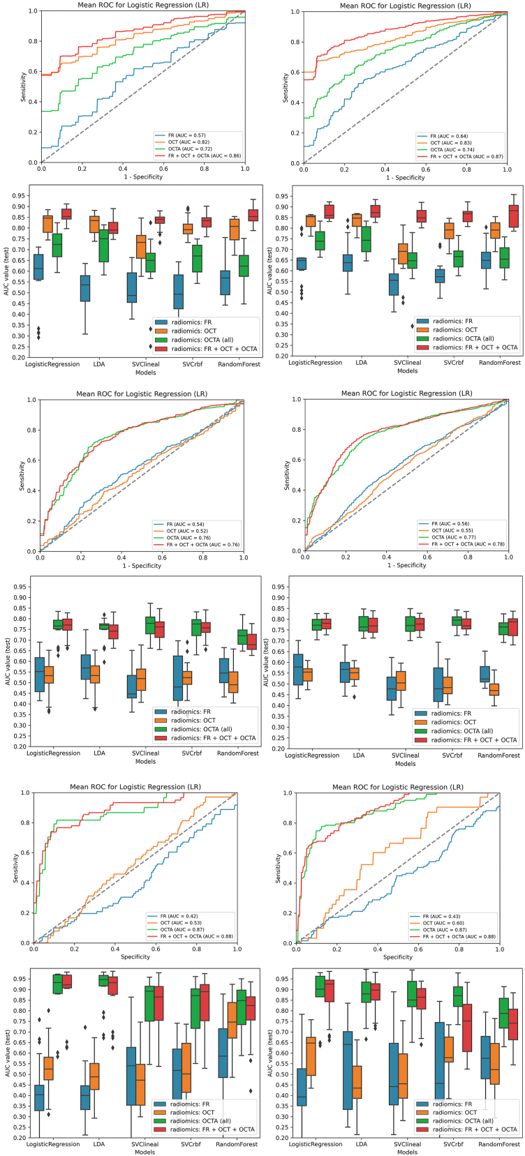
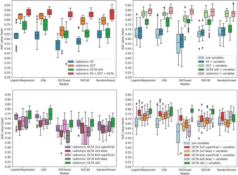
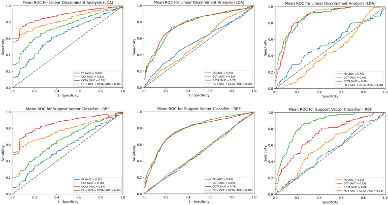
Area under the receiver operating characteristic curve (AUC) mean and standard deviation for each ML model and each individual and combined image types.
A dataset of 726 eyes (439 individuals) were included. For DM diagnosis, the greatest AUC was observed for OCT (0.82, 0.03). For DR detection, the greatest AUC was observed for OCTA (0.77, 0.03), especially in the 3 × 3 mm superficial capillary plexus OCTA scan (0.76, 0.04). For R-DR diagnosis, the greatest AUC was observed for OCTA (0.87, 0.12) and the deep capillary plexus OCTA scan (0.86, 0.08). The addition of clinical variables (age, sex, etc.) improved most models AUC for DM, DR and R-DR diagnosis. The performance of the models was similar in unilateral and bilateral eyes image datasets.
Radiomics extracted from OCT and OCTA images allow identification of patients with DM, DR, and R-DR using standard ML classifiers. OCT was the best test for DM diagnosis, OCTA for DR and R-DR diagnosis and the addition of clinical variables improved most models. This pioneer study demonstrates that radiomics-based ML techniques applied to OCT and OCTA images may be an option for DR screening in patients with type 1 DM.
Proprietary or commercial disclosure may be found after the references.
评估应用于从光学相干断层扫描(OCT)和光学相干断层扫描血管造影(OCTA)图像中提取的放射组学特征的机器学习(ML)技术对糖尿病(DM)、糖尿病视网膜病变(DR)和可转诊糖尿病视网膜病变(R-DR)诊断的准确性。
对先前一项前瞻性OCTA研究(ClinicalTrials.govNCT03422965)的视网膜图像数据集进行横断面分析。
纳入原研究中的1型糖尿病患者和对照组。
从每项研究眼的眼底视网膜造影、OCT和OCTA图像中提取放射组学特征。创建逻辑回归、线性判别分析、支持向量分类器(SVC)-线性、SVC-径向基函数和随机森林模型,以评估它们在所有图像类型中对DM、DR和R-DR诊断的准确性。
每个ML模型以及每种单独和组合图像类型的受试者操作特征曲线(AUC)下面积的均值和标准差。
纳入了一个包含726只眼(439名个体)的数据集。对于DM诊断,OCT观察到的AUC最大(0.82,0.03)。对于DR检测,OCTA观察到的AUC最大(0.77,0.03),尤其是在3×3mm浅层毛细血管丛OCTA扫描中(0.76,0.04)。对于R-DR诊断,OCTA观察到的AUC最大(0.87,0.12),深层毛细血管丛OCTA扫描中(0.86,0.08)。添加临床变量(年龄、性别等)可提高大多数模型对DM、DR和R-DR诊断的AUC。在单眼和双眼图像数据集中,模型的性能相似。
从OCT和OCTA图像中提取的放射组学特征可使用标准ML分类器识别DM、DR和R-DR患者。OCT是DM诊断的最佳检测方法,OCTA是DR和R-DR诊断的最佳检测方法,添加临床变量可改善大多数模型。这项开创性研究表明,应用于OCT和OCTA图像的基于放射组学的ML技术可能是1型糖尿病患者DR筛查的一种选择。
专有或商业披露信息可在参考文献之后找到。