• 文献检索
  • 文档翻译
  • 深度研究
  • 学术资讯
  • Suppr Zotero 插件Zotero 插件
  • 邀请有礼
  • 套餐&价格
  • 历史记录
应用&插件
Suppr Zotero 插件Zotero 插件浏览器插件Mac 客户端Windows 客户端微信小程序
定价
高级版会员购买积分包购买API积分包
服务
文献检索文档翻译深度研究API 文档MCP 服务
关于我们
关于 Suppr公司介绍联系我们用户协议隐私条款
关注我们

Suppr 超能文献

核心技术专利:CN118964589B侵权必究
粤ICP备2023148730 号-1Suppr @ 2026

文献检索

告别复杂PubMed语法,用中文像聊天一样搜索,搜遍4000万医学文献。AI智能推荐,让科研检索更轻松。

立即免费搜索

文件翻译

保留排版,准确专业,支持PDF/Word/PPT等文件格式,支持 12+语言互译。

免费翻译文档

深度研究

AI帮你快速写综述,25分钟生成高质量综述,智能提取关键信息,辅助科研写作。

立即免费体验

眼压升高损害视网膜神经节细胞的光适应。

Intraocular Pressure Elevation Compromises Retinal Ganglion Cell Light Adaptation.

机构信息

Department of Ophthalmology, Baylor College of Medicine, Houston, Texas, United States.

Department of Neuroscience, Baylor College of Medicine, Houston, Texas, United States.

出版信息

Invest Ophthalmol Vis Sci. 2020 Oct 1;61(12):15. doi: 10.1167/iovs.61.12.15.

DOI:10.1167/iovs.61.12.15
PMID:33064129
原文链接:https://pmc.ncbi.nlm.nih.gov/articles/PMC7571289/
Abstract

PURPOSE

Functional adaptation to ambient light is a key characteristic of retinal ganglion cells (RGCs), but little is known about how adaptation is affected by factors that are harmful to RGC health. We explored adaptation-induced changes to RGC physiology when exposed to increased intraocular pressure (IOP), a major risk factor for glaucoma.

METHODS

Wild-type mice of both sexes were subjected to 2 weeks of IOP elevation using the bead model. Retinas were assessed using a multielectrode array to record RGC responses to checkerboard white noise stimulation under both scotopic and photopic light levels. This information was used to calculate a spike-triggered average (STA) for each RGC with which to compare between lighting levels.

RESULTS

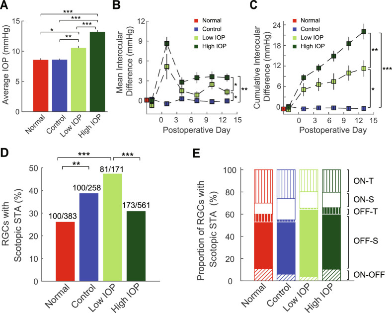
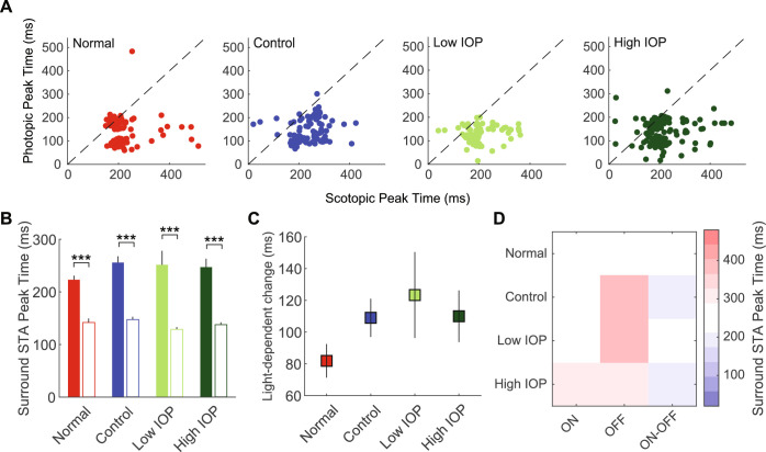
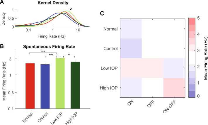
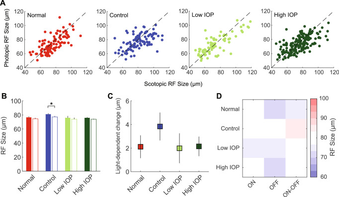
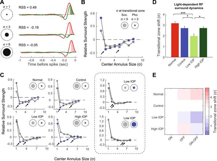
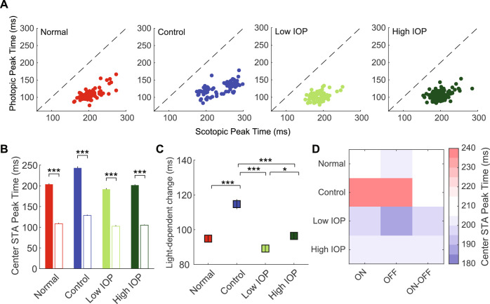
Low but not high IOP elevation resulted in several distinct RGC functional changes: (1) diminished adaptation-dependent receptive field (RF) center-surround interactions; (2) increased likelihood of a scotopic STA; and (3) increased spontaneous firing rate. Center RF size change with lighting level varied among RGCs, and both the center and surround STA peak times were consistently increased under scotopic illumination, although none of these properties were impacted by IOP level.

CONCLUSIONS

These findings provide novel evidence that RGCs exhibit reduced light-dependent adaptation and increased excitability when IOP is elevated to low but not high levels. These results may reveal functional changes that occur early in glaucoma, which can potentially be used to identify patients with glaucoma at earlier stages when intervention is most beneficial.

摘要

目的

对环境光的功能适应是视网膜神经节细胞(RGCs)的一个关键特征,但对于适应如何受到对 RGC 健康有害的因素的影响知之甚少。我们探讨了在暴露于升高的眼内压(IOP)时,即青光眼的一个主要危险因素,适应如何引起 RGC 生理学的变化。

方法

使用珠模型使雌雄两性的野生型小鼠经历 2 周的 IOP 升高。使用多电极阵列评估视网膜,以记录在暗视和明视光照水平下 RGC 对棋盘状白噪声刺激的反应。该信息用于计算每个 RGC 的尖峰触发平均(STA),以在光照水平之间进行比较。

结果

低但不是高 IOP 升高导致了几个明显的 RGC 功能变化:(1)适应依赖性感受野(RF)中心-周围相互作用减弱;(2)出现暗视 STA 的可能性增加;(3)自发放电率增加。随着光照水平的变化,中心 RF 大小在 RGC 之间变化,并且在暗视照明下,中心和周围 STA 的峰值时间始终增加,尽管这些特性都不受 IOP 水平的影响。

结论

这些发现提供了新的证据,表明当 IOP 升高到低但不是高水平时,RGC 表现出光依赖性适应的减少和兴奋性的增加。这些结果可能揭示了青光眼早期发生的功能变化,这可能有助于在干预最有益的早期阶段识别出青光眼患者。

https://cdn.ncbi.nlm.nih.gov/pmc/blobs/c126/7571289/7a5811f49dab/iovs-61-12-15-f006.jpg
https://cdn.ncbi.nlm.nih.gov/pmc/blobs/c126/7571289/7b5f0f1b1f32/iovs-61-12-15-f001.jpg
https://cdn.ncbi.nlm.nih.gov/pmc/blobs/c126/7571289/ccb8e5478433/iovs-61-12-15-f002.jpg
https://cdn.ncbi.nlm.nih.gov/pmc/blobs/c126/7571289/556138fec6c4/iovs-61-12-15-f003.jpg
https://cdn.ncbi.nlm.nih.gov/pmc/blobs/c126/7571289/cb7ef6483d84/iovs-61-12-15-f004.jpg
https://cdn.ncbi.nlm.nih.gov/pmc/blobs/c126/7571289/7d2e3256a90b/iovs-61-12-15-f005.jpg
https://cdn.ncbi.nlm.nih.gov/pmc/blobs/c126/7571289/7a5811f49dab/iovs-61-12-15-f006.jpg

相似文献

1
Intraocular Pressure Elevation Compromises Retinal Ganglion Cell Light Adaptation.眼压升高损害视网膜神经节细胞的光适应。
Invest Ophthalmol Vis Sci. 2020 Oct 1;61(12):15. doi: 10.1167/iovs.61.12.15.
2
Effects of Chronic and Acute Intraocular Pressure Elevation on Scotopic and Photopic Contrast Sensitivity in Mice.慢性和急性眼压升高对小鼠暗视和明视对比敏感度的影响。
Invest Ophthalmol Vis Sci. 2016 Jun 1;57(7):3077-87. doi: 10.1167/iovs.16-19312.
3
Mild Intraocular Pressure Elevation in Mice Reveals Distinct Retinal Ganglion Cell Functional Thresholds and Pressure-Dependent Properties.轻度眼内压升高揭示了小鼠视网膜神经节细胞功能的不同阈值和压力依赖性特征。
J Neurosci. 2019 Mar 6;39(10):1881-1891. doi: 10.1523/JNEUROSCI.2085-18.2019. Epub 2019 Jan 8.
4
Single transient intraocular pressure elevations cause prolonged retinal ganglion cell dysfunction and retinal capillary abnormalities in mice.单次短暂眼压升高可导致小鼠视网膜神经节细胞功能障碍和视网膜毛细血管异常。
Exp Eye Res. 2020 Dec;201:108296. doi: 10.1016/j.exer.2020.108296. Epub 2020 Oct 8.
5
Prolonged elevation of intraocular pressure results in retinal ganglion cell loss and abnormal retinal function in mice.眼压长期升高会导致小鼠视网膜神经节细胞丢失和视网膜功能异常。
Exp Eye Res. 2015 Jan;130:29-37. doi: 10.1016/j.exer.2014.11.007. Epub 2014 Nov 18.
6
The dark phase intraocular pressure elevation and retinal ganglion cell degeneration in a rat model of experimental glaucoma.实验性青光眼大鼠模型中的暗相眼压升高和视网膜神经节细胞变性。
Exp Eye Res. 2013 Jul;112:21-8. doi: 10.1016/j.exer.2013.04.008. Epub 2013 Apr 18.
7
Progressive degeneration of retinal and superior collicular functions in mice with sustained ocular hypertension.持续性高眼压小鼠视网膜和上丘功能的进行性退化。
Invest Ophthalmol Vis Sci. 2015 Feb 26;56(3):1971-84. doi: 10.1167/iovs.14-15691.
8
Selective Vulnerability of Specific Retinal Ganglion Cell Types and Synapses after Transient Ocular Hypertension.短暂性高眼压后特定视网膜神经节细胞类型和突触的选择性易损性
J Neurosci. 2016 Aug 31;36(35):9240-52. doi: 10.1523/JNEUROSCI.0940-16.2016.
9
Sustained ocular hypertension induces dendritic degeneration of mouse retinal ganglion cells that depends on cell type and location.持续的眼内高压会导致小鼠视网膜神经节细胞的树突退化,这取决于细胞类型和位置。
Invest Ophthalmol Vis Sci. 2013 Feb 7;54(2):1106-17. doi: 10.1167/iovs.12-10791.
10
Elevated IOP alters the space-time profiles in the center and surround of both ON and OFF RGCs in mouse.眼压升高改变了小鼠 ON 和 OFF RGC 中心和周围的时空分布。
Proc Natl Acad Sci U S A. 2017 Aug 15;114(33):8859-8864. doi: 10.1073/pnas.1706994114. Epub 2017 Jul 31.

引用本文的文献

1
Retinal ganglion cells undergo cell type-specific functional changes in a computational model of cone-mediated retinal degeneration.在锥体介导的视网膜变性计算模型中,视网膜神经节细胞会发生细胞类型特异性的功能变化。
Front Neurosci. 2023 May 18;17:1147729. doi: 10.3389/fnins.2023.1147729. eCollection 2023.
2
Selective vulnerability of the intermediate retinal capillary plexus precedes retinal ganglion cell loss in ocular hypertension.在高眼压症中,视网膜中间毛细血管丛的选择性易损性先于视网膜神经节细胞丢失。
Front Cell Neurosci. 2022 Dec 5;16:1073786. doi: 10.3389/fncel.2022.1073786. eCollection 2022.
3
Axon hyperexcitability in the contralateral projection following unilateral optic nerve crush in mice.

本文引用的文献

1
Genetic dissection of rod and cone pathways mediating light responses and receptive fields of ganglion cells in the mouse retina.遗传剖析介导光反应和小鼠视网膜神经节细胞感受野的视杆和视锥通路。
Vision Res. 2020 Feb;167:15-23. doi: 10.1016/j.visres.2019.12.005. Epub 2019 Dec 27.
2
Transcriptomic profiles of retinal ganglion cells are defined by the magnitude of intraocular pressure elevation in adult mice.成年小鼠眼内压升高的幅度决定了视网膜神经节细胞的转录组特征。
Sci Rep. 2019 Feb 22;9(1):2594. doi: 10.1038/s41598-019-39141-1.
3
Mild Intraocular Pressure Elevation in Mice Reveals Distinct Retinal Ganglion Cell Functional Thresholds and Pressure-Dependent Properties.
小鼠单侧视神经挤压后对侧投射中的轴突过度兴奋性
Brain Commun. 2022 Oct 3;4(5):fcac251. doi: 10.1093/braincomms/fcac251. eCollection 2022.
4
Abnormal perception of pattern-induced flicker colors in subjects with glaucoma.青光眼患者对模式诱导闪烁颜色的异常感知。
J Vis. 2022 Feb 1;22(2):5. doi: 10.1167/jov.22.2.5.
5
Prediction of glaucoma severity using parameters from the electroretinogram.利用视网膜电图参数预测青光眼严重程度。
Sci Rep. 2021 Dec 13;11(1):23886. doi: 10.1038/s41598-021-03421-6.
6
Soluble tumor necrosis factor-alpha-induced hyperexcitability contributes to retinal ganglion cell apoptosis by enhancing Nav1.6 in experimental glaucoma.可溶性肿瘤坏死因子-α诱导的超兴奋性通过增强实验性青光眼中的 Nav1.6 导致视网膜神经节细胞凋亡。
J Neuroinflammation. 2021 Aug 21;18(1):182. doi: 10.1186/s12974-021-02236-6.
7
Adaptive responses to neurodegenerative stress in glaucoma.青光眼神经退行性应激的适应性反应。
Prog Retin Eye Res. 2021 Sep;84:100953. doi: 10.1016/j.preteyeres.2021.100953. Epub 2021 Feb 25.
轻度眼内压升高揭示了小鼠视网膜神经节细胞功能的不同阈值和压力依赖性特征。
J Neurosci. 2019 Mar 6;39(10):1881-1891. doi: 10.1523/JNEUROSCI.2085-18.2019. Epub 2019 Jan 8.
4
Pathway-Specific Asymmetries between ON and OFF Visual Signals.视觉信号 ON 和 OFF 之间的通路特异性不对称性。
J Neurosci. 2018 Nov 7;38(45):9728-9740. doi: 10.1523/JNEUROSCI.2008-18.2018. Epub 2018 Sep 24.
5
Receptive field center-surround interactions mediate context-dependent spatial contrast encoding in the retina.感受野中心-周围相互作用介导视网膜中依赖上下文的空间对比度编码。
Elife. 2018 Sep 6;7:e38841. doi: 10.7554/eLife.38841.
6
Visual Performance as a Function of Luminance in Glaucoma: The De Vries-Rose, Weber's, and Ferry-Porter's Law.青光眼的明度视觉表现:德弗里斯-罗斯、韦伯和费里-波特定律。
Invest Ophthalmol Vis Sci. 2018 Jul 2;59(8):3416-3423. doi: 10.1167/iovs.17-22497.
7
Visual complaints of patients with glaucoma and controls under optimal and extreme luminance conditions.青光眼患者和对照者在最佳和极端亮度条件下的视觉主诉。
Acta Ophthalmol. 2018 May;96(3):288-294. doi: 10.1111/aos.13695. Epub 2018 Mar 9.
8
Foveal light and dark adaptation in patients with glaucoma and healthy subjects: A case-control study.青光眼患者与健康受试者的中心光和暗适应:病例对照研究。
PLoS One. 2018 Mar 6;13(3):e0193663. doi: 10.1371/journal.pone.0193663. eCollection 2018.
9
Axogenic mechanism enhances retinal ganglion cell excitability during early progression in glaucoma.轴突发生机制增强青光眼早期进展过程中视网膜神经节细胞的兴奋性。
Proc Natl Acad Sci U S A. 2018 Mar 6;115(10):E2393-E2402. doi: 10.1073/pnas.1714888115. Epub 2018 Feb 20.
10
Flexible Neural Hardware Supports Dynamic Computations in Retina.柔性神经硬件支持视网膜中的动态计算。
Trends Neurosci. 2018 Apr;41(4):224-237. doi: 10.1016/j.tins.2018.01.009. Epub 2018 Feb 14.