Department of Biomaterials and Composites, Faculty of Materials Science and Ceramics, AGH-University of Science and Technology, Al. Mickiewicza 30, 30-059 Kraków, Poland.
Department of Cytobiology, Faculty of Pharmacy, Collegium Medicum, Jagiellonian University, ul. Medyczna 9, 30-688 Kraków, Poland.
Int J Mol Sci. 2020 Oct 13;21(20):7541. doi: 10.3390/ijms21207541.
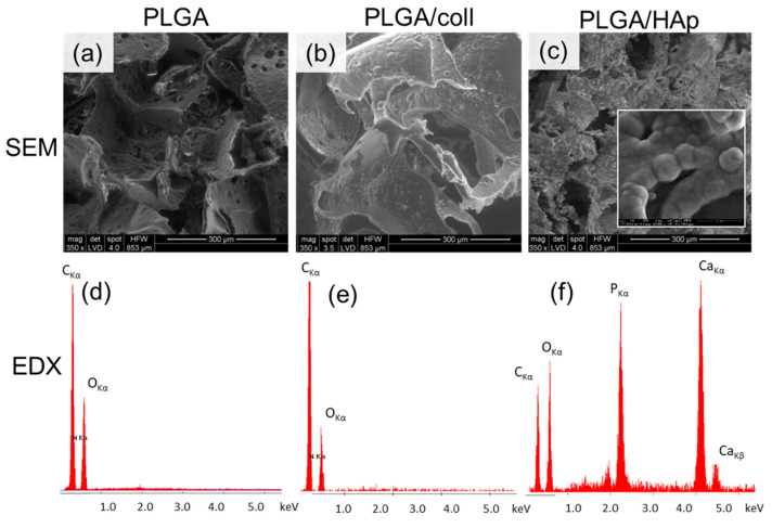
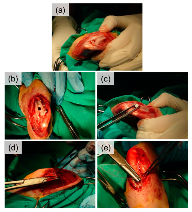
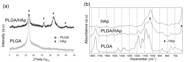
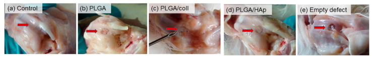
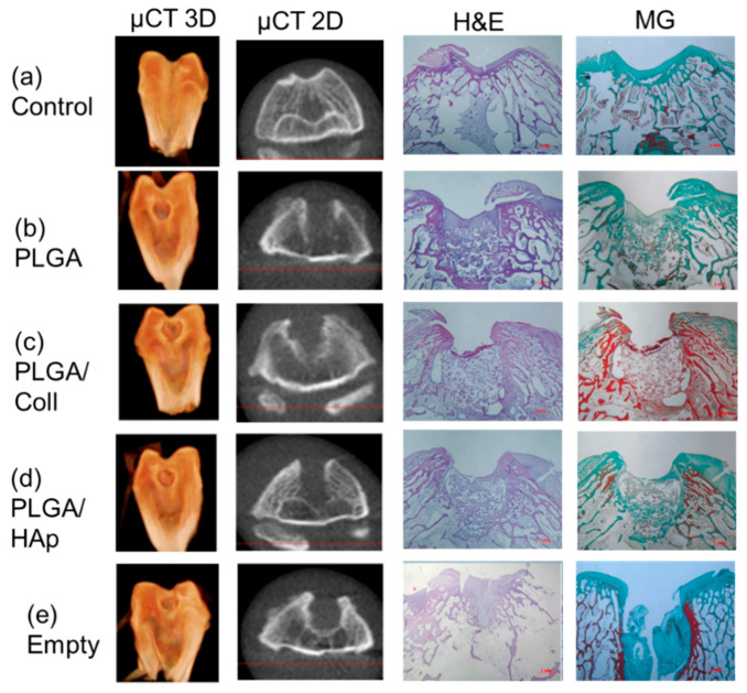
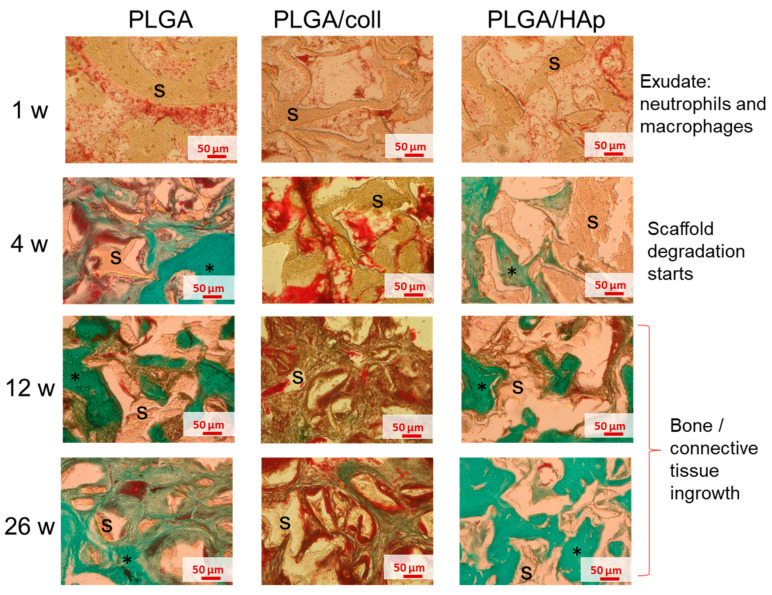
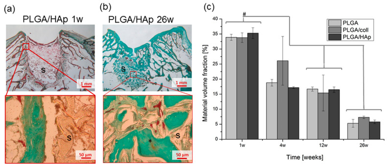
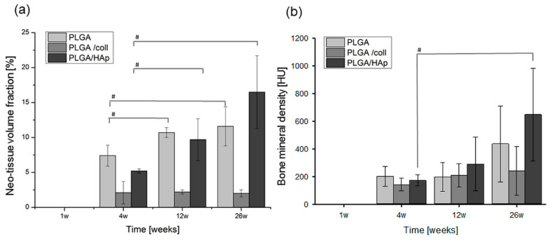
Poly(l-lactide--glycolide) (PLGA) porous scaffolds were modified with collagen type I (PLGA/coll) or hydroxyapatite (PLGA/HAp) and implanted in rabbits osteochondral defects to check their biocompatibility and bone tissue regeneration potential. The scaffolds were fabricated using solvent casting/particulate leaching method. Their total porosity was 85% and the pore size was in the range of 250-320 µm. The physico-chemical properties of the scaffolds were evaluated using scanning electron microscopy (SEM), energy dispersive X-ray spectroscopy (EDX), X-ray diffractometry (XRD), X-ray photoelectron spectroscopy (XPS), Fourier transform infrared spectroscopy (FTIR), sessile drop, and compression tests. Three types of the scaffolds (unmodified PLGA, PLGA/coll, and PLGA/HAp) were implanted into the defects created in New Zealand rabbit femoral trochlears; empty defect acted as control. Samples were extracted after 1, 4, 12, and 26 weeks from the implantation, evaluated using micro-computed tomography (µCT), and stained by Masson-Goldner and hematoxylin-eosin. The results showed that the proposed method is suitable for fabrication of highly porous PLGA scaffolds. Effective deposition of both coll and HAp was confirmed on all surfaces of the pores through the entire scaffold volume. In the in vivo model, PLGA and PLGA/HAp scaffolds enhanced tissue ingrowth as shown by histological and morphometric analyses. Bone formation was the highest for PLGA/HAp scaffolds as evidenced by µCT. Neo-tissue formation in the defect site was well correlated with degradation kinetics of the scaffold material. Interestingly, around PLGA/coll extensive inflammation and inhibited tissue healing were detected, presumably due to immunological response of the host towards collagen of bovine origin. To summarize, PLGA scaffolds modified with HAp are the most promising materials for bone tissue regeneration.
聚(L-丙交酯-乙交酯)(PLGA)多孔支架用胶原蛋白 I(PLGA/胶原)或羟基磷灰石(PLGA/HAp)进行改性,并植入兔骨软骨缺损中,以检查其生物相容性和骨组织再生潜力。支架采用溶剂浇铸/颗粒沥滤法制备。它们的总孔隙率为 85%,孔径范围为 250-320µm。支架的物理化学性质通过扫描电子显微镜(SEM)、能谱(EDX)、X 射线衍射(XRD)、X 射线光电子能谱(XPS)、傅里叶变换红外光谱(FTIR)、液滴接触角和压缩试验进行评估。将三种类型的支架(未改性的 PLGA、PLGA/胶原和 PLGA/HAp)植入新西兰兔股骨滑车的缺损中;空缺陷作为对照。植入后 1、4、12 和 26 周从植入物中提取样品,使用微计算机断层扫描(µCT)进行评估,并通过 Masson-Goldner 和苏木精-伊红染色。结果表明,该方法适用于制备高多孔 PLGA 支架。通过整个支架体积,在所有孔表面均有效沉积了胶原和 HAp。在体内模型中,PLGA 和 PLGA/HAp 支架通过组织学和形态计量学分析增强了组织向内生长。PLGA/HAp 支架的骨形成最高,这也被 µCT 证实。缺损部位新组织的形成与支架材料的降解动力学密切相关。有趣的是,在 PLGA/胶原周围检测到广泛的炎症和抑制组织愈合,这可能是由于宿主对牛源胶原的免疫反应。总之,用 HAp 改性的 PLGA 支架是骨组织再生最有前途的材料。